
Page 242 - Anatomy and Physiology of Farm Animals, 8th Edition
P. 242
Physiology of the Nervous System / 227
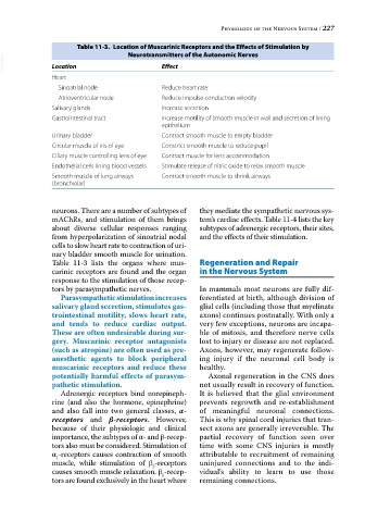
Table 11-3. Location of Muscarinic Receptors and the Effects of Stimulation by
Neurotransmitters of the Autonomic Nerves
VetBooks.ir Location Effect
Heart
Sinoatrial node Reduce heart rate
Atrioventricular node Reduce impulse conduction velocity
Salivary glands Increase secretion
Gastrointestinal tract Increase motility of smooth muscle in wall and secretion of lining
epithelium
Urinary bladder Contract smooth muscle to empty bladder
Circular muscle of iris of eye Constrict smooth muscle to reduce pupil
Ciliary muscle controlling lens of eye Contract muscle for lens accommodation
Endothelial cells lining blood vessels Stimulate release of nitric oxide to relax smooth muscle
Smooth muscle of lung airways Contract smooth muscle to shrink airways
(bronchiolar)
neurons. There are a number of subtypes of they mediate the sympathetic nervous sys
mAChRs, and stimulation of them brings tem’s cardiac effects. Table 11‐4 lists the key
about diverse cellular responses ranging subtypes of adrenergic receptors, their sites,
from hyperpolarization of sinoatrial nodal and the effects of their stimulation.
cells to slow heart rate to contraction of uri
nary bladder smooth muscle for urination.
Table 11‐3 lists the organs where mus Regeneration and Repair
carinic receptors are found and the organ in the Nervous System
response to the stimulation of those recep
tors by parasympathetic nerves. In mammals most neurons are fully dif
Parasympathetic stimulation increases ferentiated at birth, although division of
salivary gland secretion, stimulates gas- glial cells (including those that myelinate
trointestinal motility, slows heart rate, axons) continues postnatally. With only a
and tends to reduce cardiac output. very few exceptions, neurons are incapa
These are often undesirable during sur- ble of mitosis, and therefore nerve cells
gery. Muscarinic receptor antagonists lost to injury or disease are not replaced.
(such as atropine) are often used as pre- Axons, however, may regenerate follow
anesthetic agents to block peripheral ing injury if the neuronal cell body is
muscarinic receptors and reduce these healthy.
potentially harmful effects of parasym- Axonal regeneration in the CNS does
pathetic stimulation. not usually result in recovery of function.
Adrenergic receptors bind norepineph It is believed that the glial environment
rine (and also the hormone, epinephrine) prevents regrowth and re‐establishment
and also fall into two general classes, α‐ of meaningful neuronal connections.
receptors and β‐receptors. However, This is why spinal cord injuries that tran
because of their physiologic and clinical sect axons are generally irreversible. The
importance, the subtypes of α‐ and β‐recep partial recovery of function seen over
tors also must be considered. Stimulation of time with some CNS injuries is mostly
α ‐receptors causes contraction of smooth attributable to recruitment of remaining
1
muscle, while stimulation of β ‐receptors uninjured connections and to the indi
2
causes smooth muscle relaxation. β ‐recep vidual’s ability to learn to use those
1
tors are found exclusively in the heart where remaining connections.