
Page 238 - Anatomy and Physiology of Farm Animals, 8th Edition
P. 238
Physiology of the Nervous System / 223
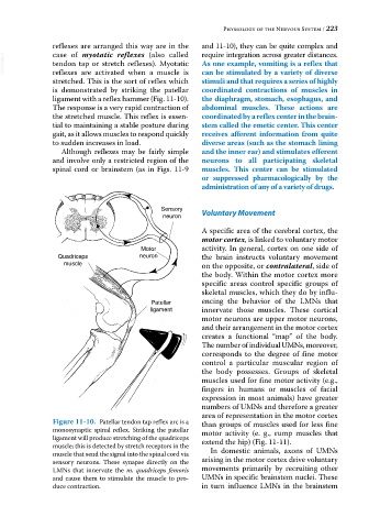
reflexes are arranged this way are in the and 11‐10), they can be quite complex and
require integration across greater distances.
case of myotatic reflexes (also called
VetBooks.ir tendon tap or stretch reflexes). Myotatic As one example, vomiting is a reflex that
reflexes are activated when a muscle is
stretched. This is the sort of reflex which can be stimulated by a variety of diverse
stimuli and that requires a series of highly
is demonstrated by striking the patellar coordinated contractions of muscles in
ligament with a reflex hammer (Fig. 11‐10). the diaphragm, stomach, esophagus, and
The response is a very rapid contraction of abdominal muscles. These actions are
the stretched muscle. This reflex is essen coordinated by a reflex center in the brain-
tial to maintaining a stable posture during stem called the emetic center. This center
gait, as it allows muscles to respond quickly receives afferent information from quite
to sudden increases in load. diverse areas (such as the stomach lining
Although reflexes may be fairly simple and the inner ear) and stimulates efferent
and involve only a restricted region of the neurons to all participating skeletal
spinal cord or brainstem (as in Figs. 11‐9 muscles. This center can be stimulated
or suppressed pharmacologically by the
administration of any of a variety of drugs.
Sensory Voluntary Movement
neuron
A specific area of the cerebral cortex, the
motor cortex, is linked to voluntary motor
Motor activity. In general, cortex on one side of
Quadriceps neuron the brain instructs voluntary movement
muscle on the opposite, or contralateral, side of
the body. Within the motor cortex more
specific areas control specific groups of
skeletal muscles, which they do by influ
Patellar encing the behavior of the LMNs that
ligament innervate those muscles. These cortical
motor neurons are upper motor neurons,
and their arrangement in the motor cortex
creates a functional “map” of the body.
The number of individual UMNs, moreover,
corresponds to the degree of fine motor
control a particular muscular region of
the body possesses. Groups of skeletal
muscles used for fine motor activity (e.g.,
fingers in humans or muscles of facial
expression in most animals) have greater
numbers of UMNs and therefore a greater
area of representation in the motor cortex
Figure 11-10. Patellar tendon tap reflex arc is a than groups of muscles used for less fine
monosynaptic spinal reflex. Striking the patellar motor activity (e. g., rump muscles that
ligament will produce stretching of the quadriceps
muscle; this is detected by stretch receptors in the extend the hip) (Fig. 11‐11).
muscle that send the signal into the spinal cord via In domestic animals, axons of UMNs
sensory neurons. These synapse directly on the arising in the motor cortex drive voluntary
LMNs that innervate the m. quadriceps femoris movements primarily by recruiting other
and cause them to stimulate the muscle to pro UMNs in specific brainstem nuclei. These
duce contraction. in turn influence LMNs in the brainstem