
Page 326 - Anatomy and Physiology of Farm Animals, 8th Edition
P. 326
Body Defenses and the Immune System / 311
Lymphocyte accumulation Afferent lymphatics
Subcapsular in deep cortex
sinus
VetBooks.ir Lymphatic capillary
Postcapillary venule
Lymphatic nodule
Medullary cord
Medullary sinus Medullary trabecula
Efferent lymphatics
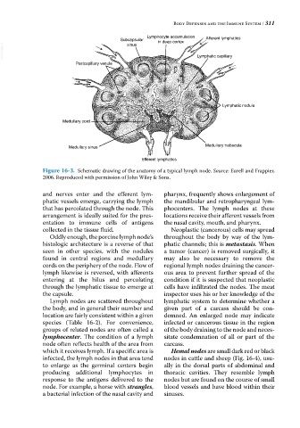
Figure 16-3. Schematic drawing of the anatomy of a typical lymph node. Source: Eurell and Frappier,
2006. Reproduced with permission of John Wiley & Sons.
and nerves enter and the efferent lym- pharynx, frequently shows enlargement of
phatic vessels emerge, carrying the lymph the mandibular and retropharyngeal lym-
that has percolated through the node. This phocenters. The lymph nodes at these
arrangement is ideally suited for the pres- locations receive their afferent vessels from
entation to immune cells of antigens the nasal cavity, mouth, and pharynx.
collected in the tissue fluid. Neoplastic (cancerous) cells may spread
Oddly enough, the porcine lymph node’s throughout the body by way of the lym-
histologic architecture is a reverse of that phatic channels; this is metastasis. When
seen in other species, with the nodules a tumor (cancer) is removed surgically, it
found in central regions and medullary may also be necessary to remove the
cords on the periphery of the node. Flow of regional lymph nodes draining the cancer-
lymph likewise is reversed, with afferents ous area to prevent further spread of the
entering at the hilus and percolating condition if it is suspected that neoplastic
through the lymphatic tissue to emerge at cells have infiltrated the nodes. The meat
the capsule. inspector uses his or her knowledge of the
Lymph nodes are scattered throughout lymphatic system to determine whether a
the body, and in general their number and given part of a carcass should be con-
location are fairly consistent within a given demned. An enlarged node may indicate
species (Table 16‐2). For convenience, infected or cancerous tissue in the region
groups of related nodes are often called a of the body draining to the node and neces-
lymphocenter. The condition of a lymph sitate condemnation of all or part of the
node often reflects health of the area from carcass.
which it receives lymph. If a specific area is Hemal nodes are small dark red or black
infected, the lymph nodes in that area tend nodes in cattle and sheep (Fig. 16‐4), usu-
to enlarge as the germinal centers begin ally in the dorsal parts of abdominal and
producing additional lymphocytes in thoracic cavities. They resemble lymph
response to the antigens delivered to the nodes but are found on the course of small
node. For example, a horse with strangles, blood vessels and have blood within their
a bacterial infection of the nasal cavity and sinuses.