
Page 331 - Anatomy and Physiology of Farm Animals, 8th Edition
P. 331
316 / Anatomy and Physiology of Farm Animals
he cardiovascular system consists of Pericardium
Tthe heart and a system of vessels for
VetBooks.ir distribution of the blood to the tissues of the The heart is partially surrounded by a
body and to the lungs for exchange of gases
(Fig. 17‐1). Regardless of whether or not the serous membrane called the pericardium.
The pericardium, like other serous tissues
blood is oxygenated, vessels that carry blood (the pleura and peritoneum), creates a
away from the heart are called arteries, and
vessels that carry blood toward the heart are
called veins. Circulation to the lungs (pul-
monary circulation) is functionally and
anatomically separate from circulation to
the rest of the body (systemic circulation).
Conceptually, it is therefore useful to regard
the heart as two separate pumps housed
within the same organ; one is a low‐pressure Right heart Left heart
pump that directs blood returning from the
body to the lungs (i.e., the pulmonary circu-
lation), and the other is a high‐pressure Parietal
pump that distributes blood to the systemic pericardium
circulation. Pericardial
cavity
Visceral
Heart pericardium
The heart is a cone‐shaped hollow muscu-
lar structure. The base is directed dorsad
to cranio‐dorsad and is attached to other
thoracic structures by large arteries, veins,
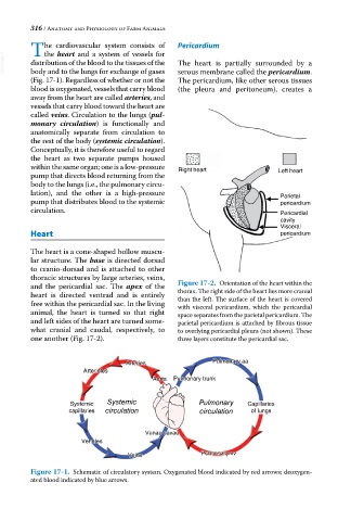
and the pericardial sac. The apex of the Figure 17-2. Orientation of the heart within the
heart is directed ventrad and is entirely thorax. The right side of the heart lies more cranial
than the left. The surface of the heart is covered
free within the pericardial sac. In the living with visceral pericardium, which the pericardial
animal, the heart is turned so that right space separates from the parietal pericardium. The
and left sides of the heart are turned some- parietal pericardium is attached by fibrous tissue
what cranial and caudal, respectively, to to overlying pericardial pleura (not shown). These
one another (Fig. 17‐2). three layers constitute the pericardial sac.
Arteries Pulmonary aa
Arterioles
Aorta Pulmonary trunk
Systemic Systemic Pulmonary Capillaries
capillaries circulation circulation of lungs
Venae cavae
Venules
Veins Pulmonary vv
Figure 17-1. Schematic of circulatory system. Oxygenated blood indicated by red arrows; deoxygen-
ated blood indicated by blue arrows.