
Page 336 - Anatomy and Physiology of Farm Animals, 8th Edition
P. 336
Anatomy of the Cardiovascular System / 321
blood is delivered into the pulmonary sys- of deoxygenated blood to the bright red of
oxygenated blood. In the adult the pulmo-
tem by contraction of the right ventricle.
VetBooks.ir After passing into the pulmonary trunk, nary circulation is the only place where
deoxygenated blood is found in arteries
blood enters the right pulmonary artery to
go to the right lung and the left pulmonary (which, by definition, carry blood away from
artery to go to the left lung. Each pulmonary the heart) and oxygenated blood is found in
artery subdivides into lobar arteries going veins (returning blood to the heart).
to individual lobes of the lungs. The lobar
arteries again subdivide many times, finally
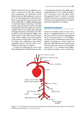
forming arterioles that supply the extensive Systemic Circulation
capillary beds of the lungs, where gaseous
exchange takes place (see Chapter 19). After Systemic circulation refers to the move-
the blood passes through the capillary bed ment of oxygenated blood to all areas of
of the lungs, it is returned to the pulmonary the body and the return of deoxygenated
veins, which usually correspond in number blood to the heart (Fig. 17‐1). The follow-
to the number of lung lobes. After leaving ing descriptions of the blood vessels of the
the lungs, these pulmonary veins deliver systemic circulation are based mainly on
oxygenated blood to the left atrium, com- the horse, although the main vessels arising
pleting the pulmonary circulation. from the aorta are common to all domestic
As gases are exchanged, the color of the species (Fig. 17‐6). Animals having differ-
blood changes from the bluish maroon color ent digestive systems, such as ruminants,
Common carotid aa. Head
Vertebral aa. Neck and head
Subclavian a.
Axillary a. Forelimb
Aorta
Foregut (+ liver) Celiac a.
Midgut Cranial mesenteric a.
Renal a. Kindney
Gonadal a.
Caudal mesenteric a. Ovary or testis
Hindgut External iliac a.
Pelvic limb Femoral a.
Internal iliac a.
Pelvic organs,
perineum
Figure 17-6. Schematic of main arteries arising from the aorta and body regions they serve. All species
will follow this basic plan. a(a)., artery/arteries.