
Page 341 - Anatomy and Physiology of Farm Animals, 8th Edition
P. 341
326 / Anatomy and Physiology of Farm Animals
VetBooks.ir
External iliac a.
Femoral a.
Popliteal a.
Cranial tibial a.
Great
metatarsal
a.
Dorsal pedal a.
Great metatarsal a. Lateral
digital
a.
Medial
digital
a.
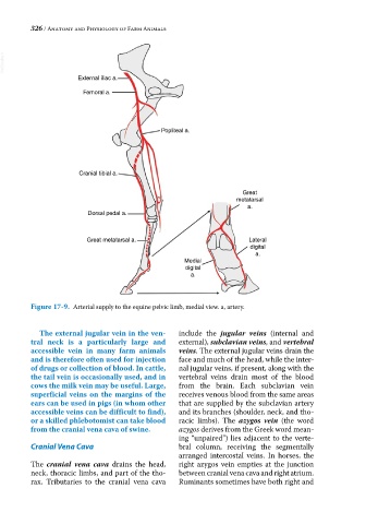
Figure 17-9. Arterial supply to the equine pelvic limb, medial view. a, artery.
The external jugular vein in the ven- include the jugular veins (internal and
tral neck is a particularly large and external), subclavian veins, and vertebral
accessible vein in many farm animals veins. The external jugular veins drain the
and is therefore often used for injection face and much of the head, while the inter-
of drugs or collection of blood. In cattle, nal jugular veins, if present, along with the
the tail vein is occasionally used, and in vertebral veins drain most of the blood
cows the milk vein may be useful. Large, from the brain. Each subclavian vein
superficial veins on the margins of the receives venous blood from the same areas
ears can be used in pigs (in whom other that are supplied by the subclavian artery
accessible veins can be difficult to find), and its branches (shoulder, neck, and tho-
or a skilled phlebotomist can take blood racic limbs). The azygos vein (the word
from the cranial vena cava of swine. azygos derives from the Greek word mean-
ing “unpaired”) lies adjacent to the verte-
Cranial Vena Cava bral column, receiving the segmentally
arranged intercostal veins. In horses, the
The cranial vena cava drains the head, right azygos vein empties at the junction
neck, thoracic limbs, and part of the tho- between cranial vena cava and right atrium.
rax. Tributaries to the cranial vena cava Ruminants sometimes have both right and