
Page 343 - Anatomy and Physiology of Farm Animals, 8th Edition
P. 343
328 / Anatomy and Physiology of Farm Animals
between the two atria forms as a flutter bypasses the pulmonary arteries through
the ductus arteriosus, a substantial ves-
valve. This unidirectional opening is called
VetBooks.ir the foramen ovale, and its structure is sel which connects the pulmonary trunk
to the aorta. In the fetus, the pressures in
such that the blood entering the right
atrium (well‐oxygenated, as much of it is the right side of the heart are greater
returning directly from the placenta) uses than those of the left side, since relatively
the one‐way flutter valve of the foramen little blood is returning from the lungs to
ovale as a convenient passageway from the the left side. As a result, the pressure is
right to the left atrium. This is one mecha- higher in the pulmonary trunk than the
nism by which blood bypasses the fetal aorta, and blood therefore flows across
lungs. the ductus arteriosus from the trunk to
Second, blood flowing from the right the aorta, bypassing the pulmonary
ventricle into the pulmonary trunk circulation.
(A) Ductus arteriosus
Fetal lungs collapsed
Foramen ovale
Caudal vena cava
Descending
aorta
Liver
Ductus venosus
Portal vein
Umbilical vein
Placenta Umbilical arteries
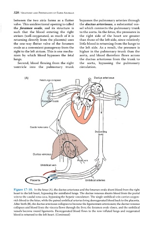
Figure 17-10. In the fetus (A), the ductus arteriosus and the foramen ovale shunt blood from the right
heart to the left heart, bypassing the uninflated lungs. The ductus venosus shunts blood from the portal
vein to the caudal vena cava, bypassing the hepatic vasculature. The single umbilical vein carries oxygen‐
rich blood to the fetus, while the paired umbilical arteries bring deoxygenated blood back to the placenta.
After birth (B), the ductus arteriosus collapses to become the ligamentum arteriosum; the ductus venosus
collapses and blood from the viscera flows through the liver, the foramen ovale closes, and the umbilical
vessels become round ligaments. Deoxygenated blood flows to the now inflated lungs and oxygenated
blood is returned to the left heart. (Continued)