
Page 347 - Anatomy and Physiology of Farm Animals, 8th Edition
P. 347
332 / Anatomy and Physiology of Farm Animals
Basic Design and Function with the parts of the lungs where the
of the Cardiovascular System
exchange of gases takes place. From the
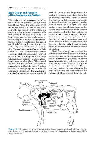
VetBooks.ir The cardiovascular system consists of the pulmonary circulation, blood re‐enters
the heart on the left side, and from here it
heart and the many vessels through which is pumped out into the systemic circula
blood flows. While the actual anatomy of tion to begin the loop again. The loop
the system makes it difficult to appreciate design means that all components of the
easily, the basic design of the system is a system must function together in a highly
continuous loop of branching vessels with coordinated and integrated fashion to
two pumps in the loop (Fig. 18‐1). The maintain blood flow throughout the sys
loop design can be best understood by tem. For example, if the right side of the
tracing the path of a single erythrocyte as it heart cannot pump an adequate amount of
travels. When the erythrocyte is pumped blood into the pulmonary circulation, the
out of the left side of the heart, it enters the left side of the heart will not receive enough
aorta and passes into the systemic circula blood to maintain flow into the systemic
tion. The systemic circulation is a subdi circulation.
vision of the cardiovascular system Blood flows through the vessels of the
consisting of all vessels associated with all cardiovascular system because of a driving
organs other than the parts of the lungs force generated by the contraction of the
where exchange of gases – oxygen and car heart. Hydrostatic pressure, or mean
bon dioxide – takes place. When blood blood pressure, in vessels is a measure of
returns from the systemic circulation, it this driving force (Chapter 2 explains
enters the right side of the heart. The right hydrostatic pressure). As the blood leaves
side of the heart pumps blood into the the heart during contraction (systole), the
pulmonary circulation. The pulmonary wall of the aorta can accommodate the
circulation consists of vessels associated volume of blood ejected from the left
Brachiocephalic
trunk Abdominal
aorta
Thoracic
Head aorta Abdomen and
and Kidney hind limbs
forelimbs Spleen
Stomach
Intestines
Renal
vein
Cranial Hepatic
vena cava artery Liver
Liver
Hepatic
Right atrium veins
Caudal vena cava
Left ventricle
Figure 18-1. General design of cardiovascular system illustrating the systemic and pulmonary circula
tions. Pulmonary circulation is shown in black. Source: Reece, 1997. Reproduced with permission of John
Wiley & Sons, Inc.