
Page 350 - Anatomy and Physiology of Farm Animals, 8th Edition
P. 350
Physiology of the Heart and Circulation / 335
Isovolumic
relaxation
VetBooks.ir contraction Ejection Rapid inflow Atrial systole
Isovolumic
Diastasis
120 Aortic Aortic valve
valve closes
100 opens Aortic pressure
Pressure (mmHg) 60 A-V valve A-V valve
80
opens
40
20 closes Atrial pressure
a c
0 Ventricular pressure
130
Volume (mL) 90 R Ventricular volume
50
P
T Electrocardiogram
Q
1st 2nd 3rd S
Phonocardiogram
Systole Diastole Systole
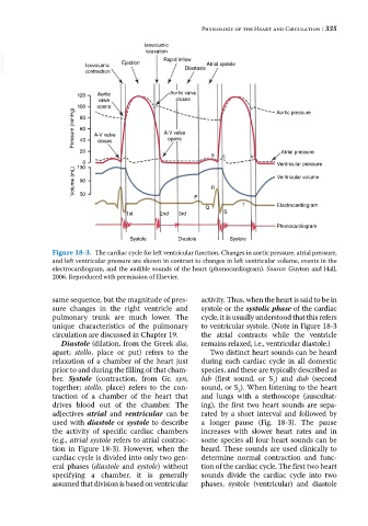
Figure 18-3. The cardiac cycle for left ventricular function. Changes in aortic pressure, atrial pressure,
and left ventricular pressure are shown in contrast to changes in left ventricular volume, events in the
electrocardiogram, and the audible sounds of the heart (phonocardiogram). Source: Guyton and Hall,
2006. Reproduced with permission of Elsevier.
same sequence, but the magnitude of pres activity. Thus, when the heart is said to be in
sure changes in the right ventricle and systole or the systolic phase of the cardiac
pulmonary trunk are much lower. The cycle, it is usually understood that this refers
unique characteristics of the pulmonary to ventricular systole. (Note in Figure 18‐3
circulation are discussed in Chapter 19. the atrial contracts while the ventricle
Diastole (dilation, from the Greek dia, remains relaxed, i.e., ventricular diastole.)
apart; stello, place or put) refers to the Two distinct heart sounds can be heard
relaxation of a chamber of the heart just during each cardiac cycle in all domestic
prior to and during the filling of that cham species, and these are typically described as
ber. Systole (contraction, from Gr. syn, lub (first sound, or S ) and dub (second
1
together; stello, place) refers to the con sound, or S ). When listening to the heart
2
traction of a chamber of the heart that and lungs with a stethoscope (auscultat
drives blood out of the chamber. The ing), the first two heart sounds are sepa
adjectives atrial and ventricular can be rated by a short interval and followed by
used with diastole or systole to describe a longer pause (Fig. 18‐3). The pause
the activity of specific cardiac chambers increases with slower heart rates and in
(e.g., atrial systole refers to atrial contrac some species all four heart sounds can be
tion in Figure 18‐3). However, when the heard. These sounds are used clinically to
cardiac cycle is divided into only two gen determine normal contraction and func
eral phases (diastole and systole) without tion of the cardiac cycle. The first two heart
specifying a chamber, it is generally sounds divide the cardiac cycle into two
assumed that division is based on ventricular phases, systole (ventricular) and diastole