
Page 355 - Anatomy and Physiology of Farm Animals, 8th Edition
P. 355
340 / Anatomy and Physiology of Farm Animals
cardiac arrhythmias in certain conditions. a
VetBooks.ir For example, it is fairly common for rac- Norepinephrine
ing Thoroughbreds to have an apparent
abnormality in A‐V node conduction at
rest. This is characterized by a reduction b
in or inhibition of the conduction of
action potentials through the A‐V node.
These disappear with exercise. A likely Stroke volume
cause is a relatively high parasympa-
thetic neural input to the heart at rest
that damps A‐V node conduction. The
relatively high parasympathetic input is
normally reduced with exercise.
Cardiac Output and Its Regulation End diastolic volume
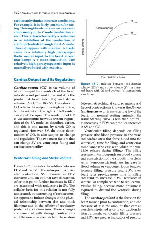
Figure 18-7. Relation between end‐diastolic
Cardiac output (CO) is the volume of volume (EDV) and stroke volume (SV) in a nor
blood pumped by a ventricle of the heart mal heart with (a) and without (b) sympathetic
into its vessel per unit time, and it is the stimulation.
product of heart rate (HR) and stroke
volume (SV): CO = HR × SV. The values for between stretching of cardiac muscle and
CO refer to the output of a single ventricle, force of contraction is known as the Frank‐
but the outputs of the right and left ventri Starling curve or Frank‐Starling law of the
cles should be equal. The regulation of HR heart. In normal resting animals, the
is via autonomic nervous system regula Frank‐Starling curve is less than optimal,
tion of the SA node, as described earlier, so increases in EDV can produce increases
and this is one means by which CO is in SV and CO.
regulated. However, SV, the other deter Ventricular filling depends on filling
minant of CO, is also subject to change pressure (the blood pressure in the veins
and regulation. The two major factors that and cardiac atria that force blood into the
can change SV are ventricular filling and ventricles), time for filling, and ventricular
cardiac contractility. compliance (the ease with which the ven
tricle relaxes during filling). The filling
pressure in turn depends on blood volume
Ventricular Filling and Stroke Volume and constriction of the smooth muscle in
veins (venoconstriction). An increase in
Figure 18‐7 illustrates the relation between blood volume or venoconstriction tends to
EDV and the SV of the subsequent ventric increase filling pressure and EDV. Slow
ular contraction. SV increases as EDV heart rates provide more time for filling
increases until an optimal EDV is reached. and tend to increase EDV. Decreases in
After that point, further increases in EDV ventricular compliance tend to reduce ven
are associated with reductions in SV. The tricular filling, because more pressure is
cellular basis for this relation is not fully required to distend the ventricle during
understood, but stretching of cardiac mus filling.
cle appears to induce changes in the physi The cardiac preload is the force on the
cal relationship between thin and thick heart muscle prior to contraction, and one
filaments and in the affinity of regulatory measure of it is the amount that cardiac
proteins for calcium ions. These changes muscle is stretched prior to contraction. In
are associated with stronger contractions intact animals, ventricular filling pressure
until the muscle is overstretched. The relation and EDV are used as indicators of preload.