
Page 358 - Anatomy and Physiology of Farm Animals, 8th Edition
P. 358
Physiology of the Heart and Circulation / 343
exchange is by simple diffusion (i.e., sub contribute to edema, an abnormal amount
or collection of fluid in the interstitial
stances move down their concentration
VetBooks.ir gradients). Gases (oxygen and carbon diox space. The primary factor that forces fluid
out of a capillary into the interstitial space
ide) and other lipid‐soluble substances
freely diffuse through capillary walls, but is the blood pressure in the capillary. The
substances that are not lipid soluble, such primary force that tends to keep fluid in
as glucose, must diffuse through pores in capillaries is the effective osmotic force
the capillary wall. Exchange by diffusion (pressure) generated by plasma proteins,
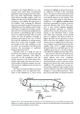
does not necessarily require the movement primarily albumin (Fig. 18‐9). This pres
of fluid between the capillary and the inter sure is also termed oncotic pressure.
stitial space. Oxygen, for example, can dif Plasma proteins generate an effective
fuse down its concentration gradient from osmotic force because the protein concen
the plasma to metabolizing cells as blood tration in the interstitial fluid is much
flows in a capillary past the cells. As stated lower than that of plasma. At the arterial
earlier, the rate of capillary exchange is end of a capillary, the blood pressure is
primarily governed by the rate of blood higher than the oncotic pressure, so some
flow into the capillaries. In resting tissues, fluid is lost from the capillary, while at the
blood flow occurs only through a small venous end of a capillary the oncotic pres
percentage of the total capillaries at any sure is higher, so some fluid moves into the
one time. As metabolism and blood flow capillary (Fig. 18‐9). A slight imbalance
increase, the percentage of capillaries between fluid loss and fluid gain by the
being perfused increases. capillaries gives rise to a net loss and pro
Typically, there is a small net loss of vides fluid for lymph formation (Fig. 18‐9).
fluid from the plasma as it flows through Like arteries, veins have smooth muscle
most capillary networks. This fluid is in their walls, but the walls of veins are
recovered via the lymphatics and ulti much thinner and more compliant.
mately returned to the blood where lym Anatomically, the venous system is also
phatics enter large veins near the heart. A characterized by a series of one way valves
small number of plasma proteins are simi that prevent blood flow back towards the
larly lost from capillaries and returned via capillaries. The compliance of venous ves
the lymph. sels permits relatively large changes in the
The forces that govern fluid movement volume of blood in the veins with minimal
at the capillary level are important clini changes in venous blood pressure. Thus,
cally in that imbalances in these forces the venous side of the circulation functions
Arterial end
Blood Venous end
flow
Net fluid Net fluid
movement Net fluid Protein movement Blood
molecules
movement flow
Lymphatic
Figure 18-9. Microvascular fluid movements. Arrows indicate direction of net fluid movement. Note
difference in relative amounts of protein molecules in plasma and interstitial fluid. Interstitial fluid with
protein moves into lymphatics to form lymph.