
Page 354 - Anatomy and Physiology of Farm Animals, 8th Edition
P. 354
Physiology of the Heart and Circulation / 339
Highly trained athletic animals, such as on the surface of the body at specific sites,
and the recorded electrical activity reflects
racing Thoroughbreds, have relatively high
VetBooks.ir levels of parasympathetic stimulation to the summated electrical activity of the
their hearts at rest.
heart. Because of the specificity of the sites
for the placement of electrodes, the pat
Atrioventricular Node and Other terns of electrical activity associated with a
cardiac cycle are predictable, and compari
Specialized Conductive Cells sons can be made between animals. The
in the Heart
lead is a specific combination of sites
where the recording electrodes are placed
The atrioventricular node (A‐V node) and on the body. An electrocardiogram (ECG)
the common bundle, or bundle of His, are is the actual recording.
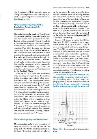
also myocardial cells specialized for con Figure 18‐6 shows a typical lead II (or
ducting action potentials. The A‐V node is limb lead II) ECG recorded from a dog.
in the intra‐atrial septum, and the common Major waves are P, Q, R, S, and T. The P
bundle extends from the A‐V node into the wave is associated with atrial depolariza
ventricle (Fig. 18‐5) through the fibrous tion. The QRS complex is associated with
connective tissue of the cardiac skeleton. ventricular depolarization, and the T wave
The cardiac skeleton separates the cardiac is associated with ventricular repolariza
muscle of the atria and ventricles, so the only tion. The period between the P and Q
direct electrical connection is through the waves is associated with A‐V node delay.
A‐V node and common bundle. The com On a typical recording from a normal dog,
mon bundle divides into several branches the waves range from 1 to 2 mV and 0.2 to
that rapidly propagate action potentials 0.3 s. Differences in wave shape and size
throughout the ventricle (Fig. 18‐5). The among species are due to normal differ
individual cells that make up these branches ences in the pattern of conduction of action
are the Purkinje fibers. potentials around the heart.
Cells of the A‐V node are pacemaker
cells, but they are specialized to conduct Arrhythmia is a general term for any
action potentials more slowly than other abnormality in cardiac electrical activ-
myocardial cells. This characteristic allows ity, including rate, rhythm, and the
enough time for the atria to depolarize com propagation of action potentials around
pletely and contract before the A‐V node the heart. Some apparently normal,
spontaneously depolarizes. The action healthy animals have a high incidence of
potentials from the SA node depolarizes the
A‐V node, which spreads the action poten R
tial into the ventricles to stimulate their
contraction. The atrial contraction com
pletes the filling of the ventricles so that
ventricular contraction can eject a larger
QRS
volume. The slow conduction through the interval
A‐V node is A‐V node delay. Sympathetic
and parasympathetic nerves that increase
and reduce conduction velocity, respec
tively, also innervate the A‐V node. P Q-T
interval
Electrocardiography and Arrhythmias
P-R (P-Q) S
interval T
Electrocardiography is the recording of Q S-T
electrical activity on the surface of the Segment
body that reflects the electrical activity in Figure 18-6. Typical canine lead II electro cardio
the heart. Recording electrodes are placed gram.