
Page 357 - Anatomy and Physiology of Farm Animals, 8th Edition
P. 357
342 / Anatomy and Physiology of Farm Animals
visualize by light microscopy. Arterioles, function as on‐off valves to regulate the
rate of blood flow from the arteries into
the smallest of arteries, are found where
VetBooks.ir arteries empty into a branching capillary capillary networks. Sympathetic vasocon-
strictor nerves innervate the smooth
network.
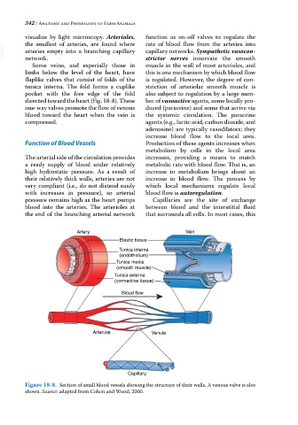
Some veins, and especially those in muscle in the wall of most arterioles, and
limbs below the level of the heart, have this is one mechanism by which blood flow
flaplike valves that consist of folds of the is regulated. However, the degree of con
tunica interna. The fold forms a cuplike striction of arteriolar smooth muscle is
pocket with the free edge of the fold also subject to regulation by a large num
directed toward the heart (Fig. 18‐8). These ber of vasoactive agents, some locally pro
one‐way valves promote the flow of venous duced (paracrine) and some that arrive via
blood toward the heart when the vein is the systemic circulation. The paracrine
compressed. agents (e.g., lactic acid, carbon dioxide, and
adenosine) are typically vasodilators; they
increase blood flow to the local area.
Function of Blood Vessels Production of these agents increases when
metabolism by cells in the local area
The arterial side of the circulation provides increases, providing a means to match
a ready supply of blood under relatively metabolic rate with blood flow. That is, an
high hydrostatic pressure. As a result of increase in metabolism brings about an
their relatively thick walls, arteries are not increase in blood flow. The process by
very compliant (i.e., do not distend easily which local mechanisms regulate local
with increases in pressure), so arterial blood flow is autoregulation.
pressure remains high as the heart pumps Capillaries are the site of exchange
blood into the arteries. The arterioles at between blood and the interstitial fluid
the end of the branching arterial network that surrounds all cells. In most cases, this
Artery Vein
Elastic tissue
Tunica interna
(endothelium)
Tunica media
(smooth muscle)
Tunica externa
(connective tissue)
Blood flow
Arteriole Venule
Capillary
Figure 18-8. Section of small blood vessels showing the structure of their walls. A venous valve is also
shown. Source: adapted from Cohen and Wood, 2000.