
Page 353 - Anatomy and Physiology of Farm Animals, 8th Edition
P. 353
338 / Anatomy and Physiology of Farm Animals
(A) (B)
Control heart rate
VetBooks.ir 30 30 (S) (PS) Threshold
Membrane potential (mV) –60 Membrane potential (mV) –60 potential
0
0
100 300
Time (ms ) Time (ms)
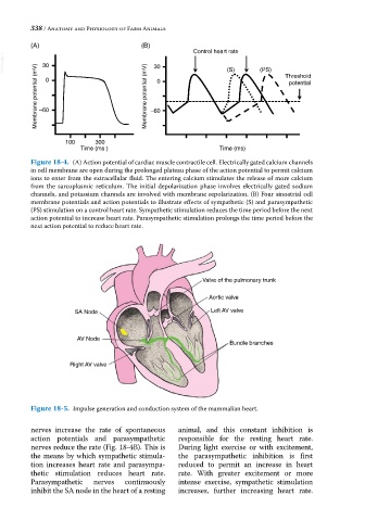
Figure 18-4. (A) Action potential of cardiac muscle contractile cell. Electrically gated calcium channels
in cell membrane are open during the prolonged plateau phase of the action potential to permit calcium
ions to enter from the extracellular fluid. The entering calcium stimulates the release of more calcium
from the sarcoplasmic reticulum. The initial depolarization phase involves electrically gated sodium
channels, and potassium channels are involved with membrane repolarization. (B) Four sinoatrial cell
membrane potentials and action potentials to illustrate effects of sympathetic (S) and parasympathetic
(PS) stimulation on a control heart rate. Sympathetic stimulation reduces the time period before the next
action potential to increase heart rate. Parasympathetic stimulation prolongs the time period before the
next action potential to reduce heart rate.
Valve of the pulmonary trunk
Aortic valve
SA Node Left AV valve
AV Node
Bundle branches
Right AV valve
Figure 18-5. Impulse generation and conduction system of the mammalian heart.
nerves increase the rate of spontaneous animal, and this constant inhibition is
action potentials and parasympathetic responsible for the resting heart rate.
nerves reduce the rate (Fig. 18‐4B). This is During light exercise or with excitement,
the means by which sympathetic stimula the parasympathetic inhibition is first
tion increases heart rate and parasympa reduced to permit an increase in heart
thetic stimulation reduces heart rate. rate. With greater excitement or more
Parasympathetic nerves continuously intense exercise, sympathetic stimulation
inhibit the SA node in the heart of a resting increases, further increasing heart rate.