
Page 344 - Anatomy and Physiology of Farm Animals, 8th Edition
P. 344
Anatomy of the Cardiovascular System / 329
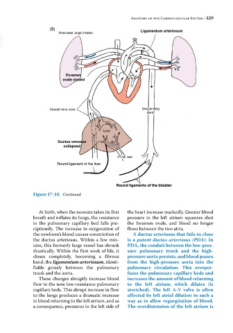
(B) Ligamentum arteriosum
Post-natal lungs inflated
VetBooks.ir
Foramen
ovale closed
Caudal vena cava Descending
aorta
Liver
Ductus venosus
collapsed
Portal vein
Round ligament of the liver
Round ligaments of the bladder
Figure 17-10. Continued
At birth, when the neonate takes its first the heart increase markedly. Greater blood
breath and inflates its lungs, the resistance pressure in the left atrium squeezes shut
in the pulmonary capillary bed falls pre- the foramen ovale, and blood no longer
cipitously. The increase in oxygenation of flows between the two atria.
the newborn’s blood causes constriction of A ductus arteriosus that fails to close
the ductus arteriosus. Within a few min- is a patent ductus arteriosus (PDA). In
utes, this formerly large vessel has shrunk PDA, the conduit between the low‐pres-
drastically. Within the first week of life, it sure pulmonary trunk and the high‐
closes completely, becoming a fibrous pressure aorta persists, and blood passes
band, the ligamentum arteriosum, identi- from the high‐pressure aorta into the
fiable grossly between the pulmonary pulmonary circulation. This overper-
trunk and the aorta. fuses the pulmonary capillary beds and
These changes abruptly increase blood increases the amount of blood returning
flow to the now low‐resistance pulmonary to the left atrium, which dilates (is
capillary beds. This abrupt increase in flow stretched). The left A‐V valve is often
to the lungs produces a dramatic increase affected by left atrial dilation in such a
in blood returning to the left atrium, and as way as to allow regurgitation of blood.
a consequence, pressures in the left side of The overdistension of the left atrium is