
Page 340 - Anatomy and Physiology of Farm Animals, 8th Edition
P. 340
Anatomy of the Cardiovascular System / 325
VetBooks.ir
Axillary a. Subscapular a.
Brachial a.
Collateral ulnar a.
Median a.
Medial
palmar a.
Medial
palmar a.
Medial
digital a.
Lateral
digital a.
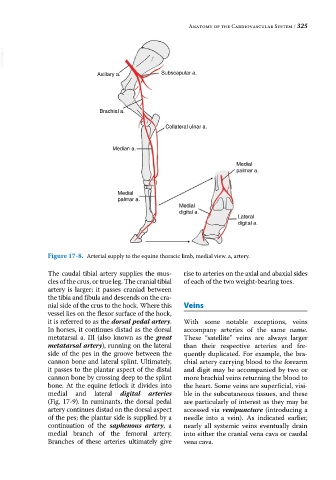
Figure 17-8. Arterial supply to the equine thoracic limb, medial view. a, artery.
The caudal tibial artery supplies the mus- rise to arteries on the axial and abaxial sides
cles of the crus, or true leg. The cranial tibial of each of the two weight‐bearing toes.
artery is larger; it passes craniad between
the tibia and fibula and descends on the cra-
nial side of the crus to the hock. Where this Veins
vessel lies on the flexor surface of the hock,
it is referred to as the dorsal pedal artery. With some notable exceptions, veins
In horses, it continues distad as the dorsal accompany arteries of the same name.
metatarsal a. III (also known as the great These “satellite” veins are always larger
metatarsal artery), running on the lateral than their respective arteries and fre-
side of the pes in the groove between the quently duplicated. For example, the bra-
cannon bone and lateral splint. Ultimately, chial artery carrying blood to the forearm
it passes to the plantar aspect of the distal and digit may be accompanied by two or
cannon bone by crossing deep to the splint more brachial veins returning the blood to
bone. At the equine fetlock it divides into the heart. Some veins are superficial, visi-
medial and lateral digital arteries ble in the subcutaneous tissues, and these
(Fig. 17‐9). In ruminants, the dorsal pedal are particularly of interest as they may be
artery continues distad on the dorsal aspect accessed via venipuncture (introducing a
of the pes; the plantar side is supplied by a needle into a vein). As indicated earlier,
continuation of the saphenous artery, a nearly all systemic veins eventually drain
medial branch of the femoral artery. into either the cranial vena cava or caudal
Branches of these arteries ultimately give vena cava.