
Page 327 - Anatomy and Physiology of Farm Animals, 8th Edition
P. 327
312 / Anatomy and Physiology of Farm Animals
Table 16-2. Selected Lymph Nodes (Lymphocenters) of Cattle
VetBooks.ir Name of Node Location
Intermandibular space
Mandibular
Parotid Rostroventral to external meatus of ear
Retropharyngeal Dorsal to pharynx
Deep cervical Dorsolateral to trachea, divided into cranial, middle, and
caudal groups
Superficial cervical (formerly prescapular) Cranial to shoulder joint
Axillary On medial aspect of shoulder near brachial plexus
Mediastinal Within the mediastinum, divided into cranial, middle,
and caudal groups
Intercostal Between ribs near thoracic vertebrae
Sternal Deep surface of sternum
Bronchial Associated with major bronchi
Lumbar Group of nodes around aorta at level of last thoracic and
first few lumbar vertebrae
Iliosacral Group of nodes around terminus of abdominal aorta
Celiac Group of nodes around origin of celiac artery
Cranial mesenteric Group of nodes around origin of cranial mesenteric artery
Subiliac (formerly prefemoral) Cranial to thigh in flank region
Superficial inguinal (scrotal or mammary) Bulls, cranial to external inguinal ring; cows, dorsocaudal
part of udder
Ischiatic Group of nodes lateral to sacrotuberous ligament
Popliteal Caudal to stifle joint
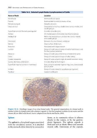
c
a
b
Figure 16-4. Histologic image of an ovine hemal node. The general organization of a hemal node is
similar to that of a lymph node with a distinct capsule (c), but the lymphatic nodules (a) are scarce and the
sinuses (b) are filled with blood. Source: adapted from Bacha and Bacha, 2012.
Spleen tissue, as in ruminants where it adheres
closely to the rumen, or by the gastros-
The spleen is a lymphoid organ associated plenic ligament. The splenic capsule is
with the circulatory system. It is attached thick and rich in elastic fibers and smooth
to the stomach either directly by connective muscle cells. Extensions of the capsule,