
Page 406 - Anatomy and Physiology of Farm Animals, 8th Edition
P. 406
Anatomy of the Digestive System / 391
Table 20-3. Species Variations in Pancreatic Ducts
VetBooks.ir Species Pancreatic Duct Accessory Pancreatic Duct
+
+
Horse
Ox Usually absent +
Pig − +
Small ruminants + −
Liver
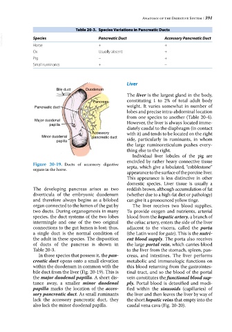
Bile duct Duodenum
The liver is the largest gland in the body,
Pancreas constituting 1 to 2% of total adult body
Pancreatic duct weight. It varies somewhat in number of
lobes and precise intra‐abdominal location
from one species to another (Table 20‐4).
Major duodenal However, the liver is always located imme
papilla
diately caudal to the diaphragm (in contact
Accessory with it) and tends to be located on the right
Minor duodenal pancreatic duct side, particularly in ruminants, in whom
papilla
the large ruminoreticulum pushes every
thing else to the right.
Individual liver lobules of the pig are
encircled by rather heavy connective tissue
Figure 20-19. Ducts of accessory digestive septa, which give a lobulated, “cobblestone”
organs in the horse.
appearance to the surface of the porcine liver.
This appearance is less distinctive in other
domestic species. Liver tissue is usually a
The developing pancreas arises as two reddish brown, although accumulation of fat
diverticula of the embryonic duodenum (whether due to a high‐fat diet or pathology)
and therefore always begins as a bilobed can give it a pronounced yellow tinge.
organ connected to the lumen of the gut by The liver receives two blood supplies.
two ducts. During organogenesis in many To provide oxygen and nutrients, arterial
species, the duct systems of the two lobes blood from the hepatic artery, a branch of
intermingle and one of the two original the celiac artery, enters the side of the liver
connections to the gut lumen is lost; thus, adjacent to the viscera, called the porta
a single duct is the normal condition of (the Latin word for gate). This is the nutri-
the adult in these species. The disposition ent blood supply. The porta also receives
of ducts of the pancreas is shown in the large portal vein, which carries blood
Table 20‐3. to the liver from the stomach, spleen, pan
In those species that possess it, the pan- creas, and intestines. The liver performs
creatic duct opens onto a small elevation metabolic and immunologic functions on
within the duodenum in common with the this blood returning from the gastrointes
bile duct from the liver (Fig. 20‐19). This is tinal tract, and so the blood of the portal
the major duodenal papilla. A short dis vein constitutes the functional blood sup-
tance away, a smaller minor duodenal ply. Portal blood is detoxified and modi
papilla marks the location of the acces- fied within the sinusoids (capillaries) of
sory pancreatic duct. As small ruminants the liver and then leaves the liver by way of
lack the accessory pancreatic duct, they the short hepatic veins that empty into the
also lack the minor duodenal papilla. caudal vena cava (Fig. 20‐20).