
Page 407 - Anatomy and Physiology of Farm Animals, 8th Edition
P. 407
392 / Anatomy and Physiology of Farm Animals
Table 20-4. Species Variations in Liver Anatomy
VetBooks.ir Species Lobation Pattern Location
Left lateral lobe
Mostly to right of midline
Swine
Left medial lobe In contact with diaphragm and ventral
Quadrate lobe body wall
Caudate lobe
Right medial lobe
Right lateral lobe
Ruminants Not grossly divided, lobes Nearly entirely to right of midline
defined by internal anatomy: In contact with diaphragm and right
Left lobe body wall
Quadrate Passes diagonally from ICS 6,
Caudate lobe caudodorsad to ICS 12
Papillary process
Caudate process
Right lobe
Equidae Partly lobated, no gall bladder Elongate shape
Left lateral lobe Approx. 3/5ths on right side midline,
Left medial lobe 2/5ths on left
Quadrate lobe In contact with diaphragm
Caudate lobe Passes caudodorsad on right body wall
from Rib 6/7 to ICS 14/15
Caudate process
Right lobe
ICS = intercostal space.
passes to the proximal duodenum into the
Hepatic artery
Viscera lumen to which it opens in common with
Portal vein the pancreatic duct on the major duodenal
papilla (see previously).
Microscopically, the morphologic unit of
the liver is the hepatic lobule, a polygonal
Hepatic lobules
Hepatic veins cylinder of liver cells (the hepatocytes) in the
Somatic structures center of which is a central vein (Fig. 20‐21).
of caudal body At the angles on the periphery, where
Caudal vena cava
adjacent hepatic lobules meet, are the portal
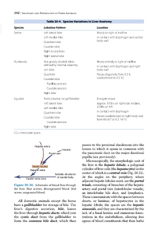
Figure 20-20. Schematic of blood flow through triads, consisting of branches of the hepatic
the liver. Blue arrows, deoxygenated blood. Red artery and portal vein (interlobular vessels),
arrow, oxygenated blood. an interlobular bile duct, and lymphatics.
These communicate with the spaces between
All domestic animals except the horse sheets, or laminae, of hepatocytes in the
have a gallbladder for storage of bile. The hepatic lobule; the spaces are the hepatic
liver’s digestive secretion, bile, leaves sinusoids, and they are characterized by the
the liver through hepatic ducts, which join lack of a basal lamina and numerous fenes
the cystic duct from the gallbladder to trations in the endothelium, allowing free
form the common bile duct, which then egress of blood constituents that then bathe