
Page 408 - Anatomy and Physiology of Farm Animals, 8th Edition
P. 408
Anatomy of the Digestive System / 393
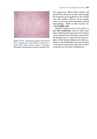
the hepatocytes. Blood (both arterial and
portal) flows from the portal canal through
VetBooks.ir c c the sinusoids and is gathered by the central
vein, the smallest tributary of the hepatic
veins. In and around the sinusoids are fixed
b b macrophages, which in this location are
called Kupffer cells.
a Between adjacent rows of liver cells is a
tiny bile canaliculus, which is little more
than a tube formed by grooves in the surfaces
c
of the apposed liver cells. Bile produced by
the hepatocytes is carried toward the peri
phery of the hepatic lobule by the bile can
Figure 20-21. Microscopic anatomy of the bovine
liver. a, hepatocytes of liver lobule; b, central vein; c, aliculi to the interlobular bile ducts located
portal veins. Source: photo courtesy of Gretchen at the portal canal (notice that this net flow
Delcambre, Colorado State University, Colorado, USA. is opposite the direction of blood flow).