
Page 68 - Clinical Small Animal Internal Medicine
P. 68
36 Section 2 Endocrine Disease
VetBooks.ir
GHRH
TRH
Hypophysiotropic GnRH
hormones
CRH NL
Others IL
AL
Pituitary hormones ACTH GH PRL TSH LH FSH MSH VP OT
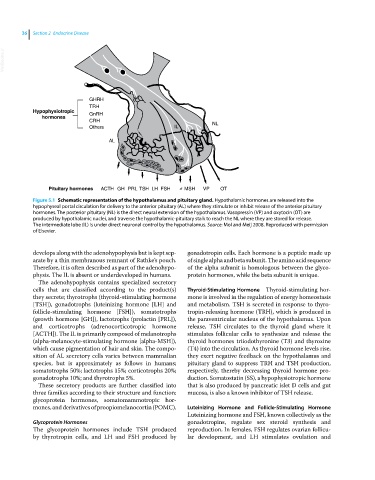
Figure 5.1 Schematic representation of the hypothalamus and pituitary gland. Hypothalamic hormones are released into the
hypophyseal portal circulation for delivery to the anterior pituitary (AL) where they stimulate or inhibit release of the anterior pituitary
hormones. The posterior pituitary (NL) is the direct neural extension of the hypothalamus. Vasopressin (VP) and oxytocin (OT) are
produced by hypothalamic nuclei, and traverse the hypothalamic‐pituitary stalk to reach the NL where they are stored for release.
The intermediate lobe (IL) is under direct neuronal control by the hypothalamus. Source: Mol and Meij 2008. Reproduced with permission
of Elsevier.
develops along with the adenohypophysis but is kept sep- gonadotropin cells. Each hormone is a peptide made up
arate by a thin membranous remnant of Rathke’s pouch. of single alpha and beta subunit. The amino acid sequence
Therefore, it is often described as part of the adenohypo- of the alpha subunit is homologous between the glyco-
physis. The IL is absent or underdeveloped in humans. protein hormones, while the beta subunit is unique.
The adenohypophysis contains specialized secretory
cells that are classified according to the product(s) Thyroid‐Stimulating Hormone Thyroid‐stimulating hor-
they secrete; thyrotrophs (thyroid‐stimulating hormone mone is involved in the regulation of energy homeostasis
[TSH]), gonadotrophs (luteinizing hormone [LH] and and metabolism. TSH is secreted in response to thyro-
follicle‐stimulating hormone [FSH]), somatotrophs tropin‐releasing hormone (TRH), which is produced in
(growth hormone [GH]), lactotrophs (prolactin [PRL]), the paraventricular nucleus of the hypothalamus. Upon
and corticotrophs (adrenocorticotropic hormone release, TSH circulates to the thyroid gland where it
[ACTH]). The IL is primarily composed of melanotrophs stimulates follicular cells to synthesize and release the
(alpha‐melanocyte‐stimulating hormone [alpha‐MSH]), thyroid hormones triiodothyronine (T3) and thyroxine
which cause pigmentation of hair and skin. The compo- (T4) into the circulation. As thyroid hormone levels rise,
sition of AL secretory cells varies between mammalian they exert negative feedback on the hypothalamus and
species, but is approximately as follows in humans; pituitary gland to suppress TRH and TSH production,
somatotrophs 50%; lactotrophs 15%; corticotrophs 20%; respectively, thereby decreasing thyroid hormone pro-
gonadotrophs 10%; and thyrotrophs 5%. duction. Somatostatin (SS), a hypophysiotropic hormone
These secretory products are further classified into that is also produced by pancreatic islet D cells and gut
three families according to their structure and function: mucosa, is also a known inhibitor of TSH release.
glycoprotein hormones, somatomammotropic hor-
mones, and derivatives of proopiomelanocortin (POMC). Luteinizing Hormone and Follicle‐Stimulating Hormone
Luteinizing hormone and FSH, known collectively as the
Glycoprotein Hormones gonadotropins, regulate sex steroid synthesis and
The glycoprotein hormones include TSH produced reproduction. In females, FSH regulates ovarian follicu-
by thyrotropin cells, and LH and FSH produced by lar development, and LH stimulates ovulation and