
Page 354 - Canine Lameness
P. 354
326 19 Stifle Region
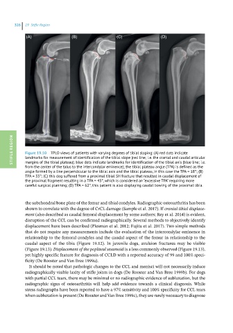
(A) (B) (C) (D)
STIFLE REGION Figure 19.10 TPLO views of patients with varying degrees of tibial sloping: (A) red dots indicate
landmarks for measurement of identification of the tibial slope (red line; i.e. the cranial and caudal articular
margins of the tibial plateau); blue dots indicate landmarks for identification of the tibial axis (blue line; i.e.
from the center of the talus to the intercondylar eminence); the tibial plateau angle (TPA) is defined as the
angle formed by a line perpendicular to the tibial axis and the tibial plateau, in this case the TPA = 18°; (B)
TPA = 33°; (C) this dog suffered from a proximal tibial SH fracture that resulted in caudal displacement of
the proximal fragment resulting in a TPA = 43°, which is considered an “excessive TPA” requiring more
careful surgical planning; (D) TPA = 62°, this patient is also displaying caudal bowing of the proximal tibia.
the subchondral bone plate of the femur and tibial condyles. Radiographic osteoarthritis has been
shown to correlate with the degree of CrCL damage (Sample et al. 2017). If cranial tibial displace-
ment (also described as caudal femoral displacement by some authors; Rey et al. 2014) is evident,
disruption of the CCL can be confirmed radiographically. Several methods to objectively identify
displacement have been described (Plesman et al. 2012; Fujita et al. 2017). Two simple methods
that do not require any measurements include the evaluation of the intercondylar eminence in
relationship to the femoral condyles and the caudal aspect of the femur in relationship to the
caudal aspect of the tibia (Figure 19.12). In juvenile dogs, avulsion fractures may be visible
(Figure 19.13). Displacement of the popliteal sesamoid is a less commonly observed (Figure 19.13),
yet highly specific feature for diagnosis of CCLD with a reported accuracy of 99 and 100% speci-
ficity (De Rooster and Van Bree 1999a).
It should be noted that pathologic changes to the CCL and menisci will not necessarily induce
radiographically visible laxity of stifle joints in dogs (De Rooster and Van Bree 1999b). For dogs
with partial CCL tears, there may be minimal or no radiographic evidence of subluxation, but the
radiographic signs of osteoarthritis will help add evidence towards a clinical diagnosis. While
stress radiographs have been reported to have a 97% sensitivity and 100% specificity for CCL tears
when subluxation is present (De Rooster and Van Bree 1999c), they are rarely necessary to diagnose