
Page 356 - Canine Lameness
P. 356
328 19 Stifle Region
(A) (C) (E) (G)
(B) (D) (F) (H)
STIFLE REGION
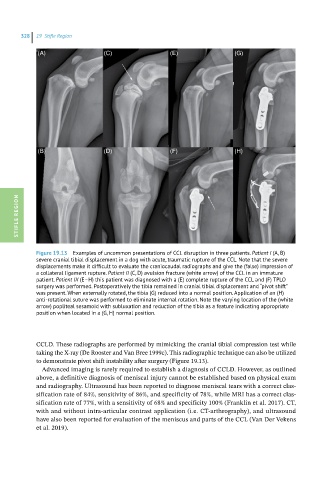
Figure 19.13 Examples of uncommon presentations of CCL disruption in three patients. Patient I (A, B)
severe cranial tibial displacement in a dog with acute, traumatic rupture of the CCL. Note that the severe
displacements make it difficult to evaluate the craniocaudal radiographs and give the (false) impression of
a collateral ligament rupture. Patient II (C, D) avulsion fracture (white arrow) of the CCL in an immature
patient. Patient III (E–H) this patient was diagnosed with a (E) complete rupture of the CCL and (F) TPLO
surgery was performed. Postoperatively the tibia remained in cranial tibial displacement and “pivot shift”
was present. When externally rotated, the tibia (G) reduced into a normal position. Application of an (H)
anti-rotational suture was performed to eliminate internal rotation. Note the varying location of the (white
arrow) popliteal sesamoid with subluxation and reduction of the tibia as a feature indicating appropriate
position when located in a (G, H) normal position.
CCLD. These radiographs are performed by mimicking the cranial tibial compression test while
taking the X-ray (De Rooster and Van Bree 1999c). This radiographic technique can also be utilized
to demonstrate pivot shift instability after surgery (Figure 19.13).
Advanced imaging is rarely required to establish a diagnosis of CCLD. However, as outlined
above, a definitive diagnosis of meniscal injury cannot be established based on physical exam
and radiography. Ultrasound has been reported to diagnose meniscal tears with a correct clas -
sification rate of 84%, sensitivity of 86%, and specificity of 78%, while MRI has a correct clas -
sification rate of 77%, with a sensitivity of 68% and specificity 100% (Franklin et al. 2017). CT,
with and without intra-articular contrast application (i.e. CT-arthrography), and ultrasound
have also been reported for evaluation of the meniscus and parts of the CCL (Van Der Vekens
et al. 2019).