
Page 355 - Canine Lameness
P. 355
19.4 Cranial Cruciate igament Disease 327
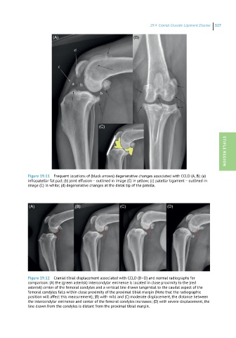
(A) (B)
(C)
Figure 19.11 Frequent locations of (black arrows) degenerative changes associated with CCLD (A, B): (a) STIFLE REGION
infrapatellar fat pad; (b) joint effusion – outlined in image (C) in yellow; (c) patellar ligament – outlined in
image (C) in white; (d) degenerative changes at the distal tip of the patella.
(A) (B) (C) (D)
Figure 19.12 Cranial tibial displacement associated with CCLD (B–D) and normal radiographs for
comparison: (A) the (green asterisk) intercondylar eminence is located in close proximity to the (red
asterisk) center of the femoral condyles and a vertical line drawn tangential to the caudal aspect of the
femoral condyles falls within close proximity of the proximal tibial margin (Note that the radiographic
position will affect this measurement); (B) with mild and (C) moderate displacement, the distance between
the intercondylar eminence and center of the femoral condyles increases; (D) with severe displacement, the
line drawn from the condyles is distant from the proximal tibial margin.