
Page 73 - Canine Lameness
P. 73
4.2 Neuroaoaromy NeoaNed aro Lomb eanaLra 45
(A)
(B) (C)
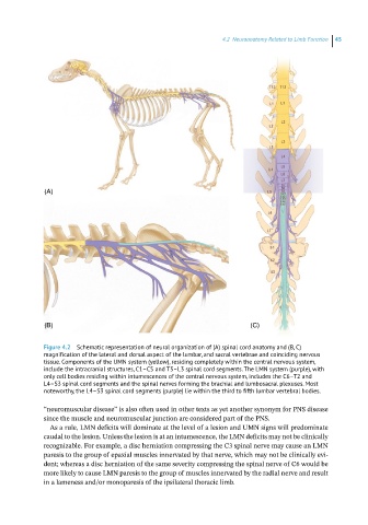
Figure 4.2 Schematic representation of neural organization of (A) spinal cord anatomy and (B, C)
magnification of the lateral and dorsal aspect of the lumbar, and sacral vertebrae and coinciding nervous
tissue. Components of the UMN system (yellow), residing completely within the central nervous system,
include the intracranial structures, C1–C5 and T3–L3 spinal cord segments. The LMN system (purple), with
only cell bodies residing within intumescences of the central nervous system, includes the C6–T2 and
L4–S3 spinal cord segments and the spinal nerves forming the brachial and lumbosacral plexuses. Most
noteworthy, the L4–S3 spinal cord segments (purple) lie within the third to fifth lumbar vertebral bodies.
“neuromuscular disease” is also often used in other texts as yet another synonym for PNS disease
since the muscle and neuromuscular junction are considered part of the PNS.
As a rule, LMN deficits will dominate at the level of a lesion and UMN signs will predominate
caudal to the lesion. Unless the lesion is at an intumescence, the LMN deficits may not be clinically
recognizable. For example, a disc herniation compressing the C3 spinal nerve may cause an LMN
paresis to the group of epaxial muscles innervated by that nerve, which may not be clinically evi-
dent; whereas a disc herniation of the same severity compressing the spinal nerve of C6 would be
more likely to cause LMN paresis to the group of muscles innervated by the radial nerve and result
in a lameness and/or monoparesis of the ipsilateral thoracic limb.