
Page 74 - Canine Lameness
P. 74
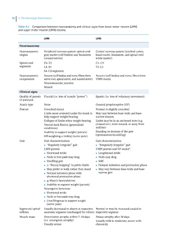
46  4  The Neurologic Examination

            Table 4.1  Comparison between neuroanatomy and clinical signs from lower motor neuron (LMN)
            and upper motor neuron (UMN) lesions.


                           LMN                            UMN
             Neuroanatomy
             Neuroanatomic   Peripheral nervous system: spinal cord   Central nervous system (cerebral cortex,
             origins       gray matter (cell bodies) and brainstem   basal nuclei, brainstem, and spinal cord
                           (cranial nerves)               white matter)
             Spinal cord   C6–T2                          C1–C5
             segments      L4–S3                          T3–L3
                           S4–Cd segments
             Neuroanatomic   Neuron (cell bodies and nerve fibers form   Neuron (cell bodies and nerve fibers form
             components    nerve root, spinal nerve, and named nerve) UMN tracts)
                           Neuromuscular junction
                           Muscle
             Clinical signs
             Quality of paresis   Flaccid (i.e. loss of muscle “power”)  Spastic (i.e. loss of voluntary movement)
             or paralysis
             Ataxia type   None                           General proprioceptive (GP)
             Posture       Crouched stance                Normal to slightly crouched
                           Limbs more centered under the trunk to  May vary between base‐wide and base‐
                           help support weight‐bearing    narrow stances
                           Collapse of limbs when weight‐bearing  Limbs may be in an awkward state (e.g.
                           Ventral neck flexion (generalized   crossed over, more toward, or away from
                           conditions)                    midline)
                           Inability to support weight (paresis)  Standing on dorsum of the paw
                           Off‐weighting a limb(s) (nerve pain)  (spontaneous knuckling)
             Gait          Gait characterization:         Gait characterization:
                              “Regularly irregular” gait     “Irregularly irregular” gait
                           ●                              ●
                                                                              a
                           LMN paresis:                   UMN paresis and GP ataxia :
                              Shortened stride               Lengthened stride
                           ●                              ●
                              Nails or foot pads may drag    Nails may drag
                           ●                              ●
                              Shuffling gait                 Stiff
                           ●                              ●
                              ± “Bunny hopping” in pelvic limbs    Delayed initiation and protraction phase
                           ●                              ●
                              May prefer to walk rather than stand    May vary between base‐wide and base‐
                           ●                              ●
                              Normal initiation phase with    narrow gait
                           ●
                             shortened protraction phase
                              ± Muscle fasciculations
                           ●
                              Inability to support weight (paresis)
                           ●
                           Neurogenic lameness:
                              Shortened stride
                           ●
                              Nails or foot pads may drag
                           ●
                              Unwillingness to support weight
                           ●
                             (nerve pain)
             Segmental spinal   Usually decreased or absent at respective  Normal or may be increased caudal to
             reflexes      anatomic segment (unchanged for others) respective segment
             Muscle mass   Denervation atrophy within 7–10 days    Disuse atrophy after 30 days
                           (i.e. neurogenic atrophy)      Usually mild to moderate; severe with
                           Usually severe                 chronicity