
Page 75 - Canine Lameness
P. 75
4.2 Neuroaoaromy NeoaNed aro Lomb eanaLra 47
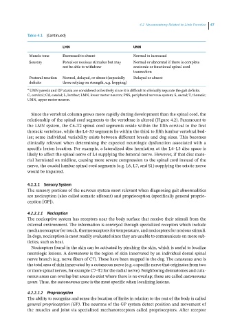
Table 4.1 (Continued)
LMN UMN
Muscle tone Decreased to absent Normal to increased
Sensory Perceives noxious stimulus but may Normal or abnormal if there is complete
not be able to withdraw anatomic or functional spinal cord
transection
Postural reaction Normal, delayed, or absent (especially Delayed or absent
deficits those relying on strength, e.g. hopping)
a UMN paresis and GP ataxia are considered collectively since it is difficult to clinically separate the gait deficits.
C, cervical; Cd, caudal; L, lumbar; LMN, lower motor neuron; PNS, peripheral nervous system; S, sacral; T, thoracic;
UMN, upper motor neuron.
Since the vertebral column grows more rapidly during development than the spinal cord, the
relationship of the spinal cord segments to the vertebrae is altered (Figure 4.2). Paramount to
the LMN system, the C6–T2 spinal cord segments reside within the fifth cervical to the first
thoracic vertebrae, while the L4–S3 segments lie within the third to fifth lumbar vertebral bod-
ies; some individual variability exists between different breeds and dog sizes. This becomes
clinically relevant when determining the expected neurologic dysfunction associated with a
specific lesion location. For example, a lateralized disc herniation at the L4–L5 disc space is
likely to affect the spinal nerve of L4 supplying the femoral nerve. However, if that disc mate -
rial herniated on midline, causing more severe compression to the spinal cord instead of the
nerve, the caudal lumbar spinal cord segments (e.g. L6, L7, and S1) supplying the sciatic nerve
would be impaired.
4.2.2.2 Sensory System
The sensory portions of the nervous system most relevant when diagnosing gait abnormalities
are nociception (also called somatic afferent) and proprioception (specifically general proprio-
ception [GP]).
4.2.2.2.1 Nociception
The nociceptive system has receptors near the body surface that receive their stimuli from the
external environment. The information is conveyed through specialized receptors which include
mechanoreceptor for touch, thermoreceptors for temperature, and nociceptors for noxious stimuli.
In dogs, nociception is most readily evaluated since they are unable to communicate on more sub-
tleties, such as heat.
Nociceptors found in the skin can be activated by pinching the skin, which is useful to localize
neurologic lesions. A dermatome is the region of skin innervated by an individual dorsal spinal
nerve branch (e.g. nerve fibers of C7). These have been mapped in the dog. The cutaneous area is
the total area of skin innervated by a cutaneous nerve (e.g. a specific nerve that originates from two
or more spinal nerves, for example C7–T2 for the radial nerve). Neighboring dermatomes and cuta-
neous areas can overlap but areas do exist where there is no overlap; these are called autonomous
zones. Thus, the autonomous zone is the most specific when localizing lesions.
4.2.2.2.2 Proprioception
The ability to recognize and sense the location of limbs in relation to the rest of the body is called
general proprioception (GP). The neurons of the GP system detect position and movement of
the muscles and joint via specialized mechanoreceptors called proprioceptors. After receptor