
Page 183 - Clinical Small Animal Internal Medicine
P. 183
16 Imaging in Cardiovascular Disease 151
VetBooks.ir (a) (a)
(b)
(b)
(c)
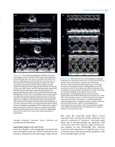
Figure 16.19 M‐mode echocardiograms obtained from two
normal dogs at the ventricular level (from the right parasternal
transventricular short‐axis view, (a)) and the mitral valve level Figure 16.20 Abnormal M‐mode echocardiograms obtained
(from the right parasternal transmitral short‐axis view, (b)). (a) from three cats with heart diseases at the ventricular level (a,b)
This ventricular M‐mode echocardiogram displays the right and the mitral valve level (c). (a) In this cat with taurine
ventricular myocardial wall (RVW) and the right ventricular deficiency‐induced dilated cardiomyopathy, the ventricular
cavity (RV) at the top of the image, the left ventricle (LV) and the M‐mode echocardiogram shows marked dilation of the left
LV free wall (LVFW) below, with the interventricular septum (IVS) ventricular cavity (LV) with almost no difference between the
between the two ventricular cavities. The M‐mode cursor is end‐diastolic and end‐systolic LV diameters. (b) This ventricular
placed perpendicular to the IVS and the LVFW between the two M‐mode echocardiogram in a Maine Coon cat with
left papillary muscles. The LV end‐diastolic (LVd) and end‐systolic hypertrophic cardiomyopathy shows severe symmetric
(LVs) diameters can be measured (double arrows), and the left hypertrophy (end‐diastolic interventricular septum and LV free
ventricular fractional shortening (expressed in percentage and wall >6 mm, double arrows). (c) This M‐mode echocardiogram
defined as the difference between the LVd and LVs divided by obtained at the mitral valve level in a cat with hypertrophic
LVd) can then be calculated. (b) This M‐mode echocardiogram at cardiomyopathy shows a significant systolic anterior motion of
the mitral valve level shows the M‐shaped motion of the anterior the mitral valve (arrows), which is characterized by an
mitral valve leaflet during diastole. The E point and the smaller A abnormal mitral valve septal contact during diastole, leading
point represent the maximum valve opening during the rapid to LV outflow tract obstruction. IVS, interventricular septum;
ventricular filling phase and the atrial contraction, respectively. LVFW, left ventricular free wall.
Closure of the mitral valve occurs after atrial contraction, at
end‐diastole.
flow across the ventricular septal defect (>5 m/s)
associated with normal peak systolic pulmonary flow
vascular resistance increases, lower velocities are velocity is indicative of “restrictive” ventricular septal
recorded across the ductus. shunt, that is, hemodynamically not significant, with
conservation of the left ventricle–right ventricle pres-
Septal Defects (Figures 16.31c and 16.31d) sure gradient (at least 100 mmHg). Flow velocities
Color flow Doppler echocardiography associated with across the atrial septal defect are typically low (<1 m/s),
spectral Doppler modes may identify small defects that as the left atrium–right atrium pressure gradient is only
cannot be visualized on 2D mode images. High‐velocity several mmHg (Figure 16.31d).