
Page 188 - Clinical Small Animal Internal Medicine
P. 188
156 Section 3 Cardiovascular Disease
VetBooks.ir (a): Dog with PAH (b): Dog with pulmonic stenosis
Notch
(c): Dog with pulmonic stenosis
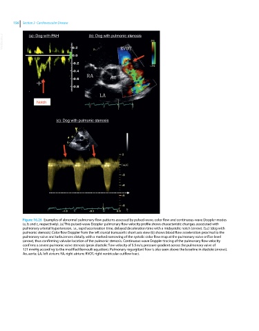
Figure 16.26 Examples of abnormal pulmonary flow patterns assessed by pulsed‐wave, color flow and continuous‐wave Doppler modes
(a, b and c, respectively). (a) This pulsed‐wave Doppler pulmonary flow velocity profile shows characteristic changes associated with
pulmonary arterial hypertension, i.e., rapid acceleration time, delayed deceleration time with a midsystolic notch (arrow). (b,c) (dog with
pulmonic stenosis) Color flow Doppler from the left cranial transaortic short axis view (b) shows blood flow acceleration proximal to the
pulmonary valve and turbulences distally, with a marked narrowing of the systolic color flow map at the pulmonary valve orifice level
(arrow), thus confirming valvular location of the pulmonic stenosis. Continuous‐wave Doppler tracing of the pulmonary flow velocity
confirms a severe pulmonic valve stenosis (peak diastolic flow velocity of 5.5 m/s; pressure gradient across the pulmonary valve of
121 mmHg according to the modified Bernoulli equation). Pulmonary regurgitant flow is also seen above the baseline in diastole (arrows).
Ao, aorta; LA, left atrium; RA, right atrium; RVOT, right ventricular outflow tract.