
Page 190 - Clinical Small Animal Internal Medicine
P. 190
158 Section 3 Cardiovascular Disease
VetBooks.ir (a)
(b) (c)
(d)
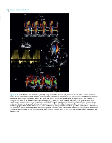
Figure 16.28 Evaluation of aortic insufficiency severity using color‐coded M‐mode (a,d), color flow (c) and continuous‐wave Doppler
modes (b). The color‐coded M‐mode from the right parasternal five‐chamber view may be used to measure the height (or cross‐sectional
area) of the regurgitant aortic jet (blue arrows) relative to the height (cross‐sectional area) of the left ventricular outflow tract (LVOT)
(double arrows, a and d). (a) shows a mild aortic insufficiency as the jet height /LVOT height is only 8% (<25%). Conversely, the aortic
insufficiency in (d) is considered as severe, as the jet height/LVOT height is high (i.e., 84%; >65%). In contrast to Figure 16.27c, a severe
aortic insufficiency extending nearly to the apex (blue arrow) is also shown in (c). Continuous‐wave Doppler mode confirms an aortic
insufficiency signal above the baseline in (b). The aortic insufficiency velocity remains stable throughout the diastolic time (white arrows).
This means that, despite the regurgitation, the pressure gradient across the aortic valve remains unchanged during diastole (no decrease
in aortic diastolic pressure), which confirms that the regurgitation is mild. Ao, aorta; LA, left atrium; LV, left ventricle; RA, right atrium; RV,
right ventricle.