
Page 191 - Clinical Small Animal Internal Medicine
P. 191
16 Imaging in Cardiovascular Disease 159
VetBooks.ir (a) (b)
(c) (d)
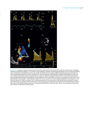
Figure 16.29 Examples of abnormal diastolic transmitral inflows obtained from left parasternal apical four‐chamber views. (a) Delayed
relaxation transmitral flow pattern in a cat, which is characterized by a delayed and prolonged early diastolic E wave (with an inverted E/A
ratio). (b) Restrictive transmitral flow pattern, with a high early diastolic E wave (owing to high left atrial pressure) and a diminished A
wave (owing to decreased left ventricular compliance) in a cat with restrictive cardiomyopathy. Doppler echocardiograms of two dogs
with congenital mitral valve stenosis are presented in (c) and (d). (c) Color flow Doppler mode across the stenotic mitral valve shows a
narrow aliased turbulent signal during diastole. (d) Spectral Doppler of the mitral inflow (continuous‐wave Doppler mode) reveals a high
peak diastolic E‐wave velocity (1.9 m/s, reference range: 0.58–1.17 m/s; see Table 16.4) and a high peak diastolic A‐wave velocity (2.1 m/s,
reference range: 0.39–0.86 m/s; see Table 16.4). The pressure half time for the E wave, which is defined as the time needed for the peak
transvalvular pressure gradient to fall to its half value, may be calculated automatically by tracing (white arrow) the deceleration slope of
the E wave. In this case, the pressure half time is markedly prolonged (290 ms, reference range: <50 ms), thus confirming a severe mitral
valve stenosis. LA, left atrium; LV, left ventricle.