
Page 195 - Clinical Small Animal Internal Medicine
P. 195
16 Imaging in Cardiovascular Disease 163
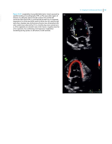
Figure 16.33 Longitudinal myocardial deformation (strain) assessed by (a)
VetBooks.ir interest, including the interventricular septum (IVS) and the left
speckle tracking echocardiography (STE). (a) The myocardial region of
ventricular free wall (LVFW), is automatically divided into six segments
(segments #1 to #3 for the LVFW, and #4 to #6 for the IVS) using the left
apical four‐chamber view. (b) Example of normal two‐dimensional (2D)
color‐coded strain obtained by STE in a healthy dog (same animal as in
a). All six segments are colored in red, which means a negative strain for
all six segments, thus confirming a homogeneous longitudinal
shortening during systole. LA, left atrium; LV, left ventricle.
(b)