
Page 198 - Clinical Small Animal Internal Medicine
P. 198
166 Section 3 Cardiovascular Disease
VetBooks.ir RA LA RA LA
– I + + aVR 0 I aVL + +
RA – – LA – –
0 0
III II III
II aVFaVF
+ +
LA
+
LL
RL LL RL LL
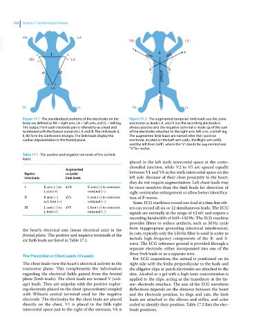
Figure 17.1 The standardized positions of the electrodes on the Figure 17.2 The augmented (unipolar) limb leads use the same
body are defined as RA = right arm, LA = left arm, and LL = left leg. electrodes as leads I, II, and III but the recording electrode is
The output from each electrode pair is referred to as a lead and always positive and the negative terminal is made up of the sum
numbered with the Roman numerals I, II, and III. The limb leads (I, of the electrodes attached to the right arm, left arm, and left leg.
II, III) form the Einthoven’s triangle. The limb leads display the The augmented limb leads are named after their positive
cardiac depolarization in the frontal plane. electrode, located on the Left arm (aVL), the Right arm (aVR),
and the left Foot (aVF), where the “a” stands for augmented and
“V” for vector.
Table 17.1 The positive and negative terminals of the six limb
leads
placed in the left sixth intercostal space at the costo-
chondral junction, while V2 to V5 are spaced equally
Augmented
Bipolar unipolar between V1 and V6 in the sixth intercostal space on the
limb leads limb leads left side. Because of their close proximity to the heart,
they do not require augmentation. Left chest leads may
I R arm (−) to aVR R arm (+) to common be more sensitive than the limb leads for detection of
L arm (+) terminal (−) right ventricular enlargement or allow better identifica-
II R arm (−) aVL L arm (+) to common tion of P‐waves.
to L foot (+) terminal (−) Some ECG machines record one lead at a time but oth-
III L arm (−) to aVF L foot (+) to common ers can record all six or 12 simultaneous leads. The ECG
L foot (+) terminal (−) signals are normally in the range of ±2 mV and require a
recording bandwidth of 0.05–150 Hz. The ECG machine
provides filters to reduce artifacts, such as 50 Hz cycle
the heart’s electrical axis (mean electrical axis) in the from inappropriate grounding (electrical interference).
frontal plane. The positive and negative terminals of the In cats, typically only the 150 Hz filter is used in order to
six limb leads are listed in Table 17.1. include high‐frequency components of the R‐ and S‐
wave. The ECG reference ground is provided through a
separate electrode, either incorporated into one of the
three limb leads or as a separate wire.
The Precordial or Chest Leads (V Leads)
For ECG acquisition, the animal is positioned on its
The chest leads view the heart’s electrical activity in the right side with the limbs perpendicular to the body and
transverse plane. This complements the information the alligator clips or patch electrodes are attached to the
regarding the electrical fields gained from the frontal skin. Alcohol or a gel with a high ionic concentration is
plane (limb leads). The chest leads are termed V (volt- applied to the clips, acting as the transducer at the tis-
age) leads. They are unipolar with the positive explor- sue–electrode interface. The size of the ECG waveform
ing electrode placed on the chest (precordium) coupled deflections depends on the distance between the heart
with Wilson’s central terminal used for the negative and the electrode position. In dogs and cats, the limb
electrode. The electrodes for the chest leads are placed leads are attached to the elbows and stifles, and color
directly on the chest. V1 is placed in the fifth right coded to identify their position. Table 17.2 lists the elec-
intercostal space just to the right of the sternum, V6 is trode positions.