
Page 202 - Clinical Small Animal Internal Medicine
P. 202
170 Section 3 Cardiovascular Disease
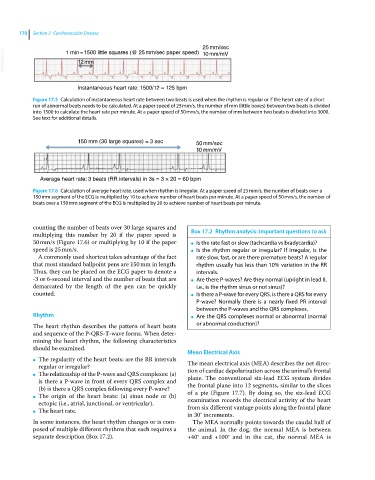
25 mm/sec
VetBooks.ir 12mm 10 mm/mV
1 min=1500 little squares (@ 25mm/sec paper speed)
Instantaneous heart rate: 1500/12 = 125 bpm
Figure 17.5 Calculation of instantaneous heart rate between two beats is used when the rhythm is regular or if the heart rate of a short
run of abnormal beats needs to be calculated. At a paper speed of 25 mm/s, the number of mm (little boxes) between two beats is divided
into 1500 to calculate the heart rate per minute. At a paper speed of 50 mm/s, the number of mm between two beats is divided into 3000.
See text for additional details.
150 mm (30 large squares) = 3 sec 50 mm/sec
10 mm/mV
Average heart rate: 3 beats (RR intervals) in 3s = 3 × 20 = 60 bpm
Figure 17.6 Calculation of average heart rate, used when rhythm is irregular. At a paper speed of 25 mm/s, the number of beats over a
150 mm segment of the ECG is multiplied by 10 to achieve number of heart beats per minute. At a paper speed of 50 mm/s, the number of
beats over a 150 mm segment of the ECG is multiplied by 20 to achieve number of heart beats per minute.
counting the number of beats over 30 large squares and Box 17.2 Rhythm analysis: important questions to ask
multiplying this number by 20 if the paper speed is
50 mm/s (Figure 17.6) or multiplying by 10 if the paper ● Is the rate fast or slow (tachcardia vs bradycardia)?
speed is 25 mm/s. ● Is the rhythm regular or irregular? If irregular, is the
A commonly used shortcut takes advantage of the fact rate slow, fast, or are there premature beats? A regular
that most standard ballpoint pens are 150 mm in length. rhythm usually has less than 10% variation in the RR
Thus, they can be placed on the ECG paper to denote a intervals.
‐3 or 6‐second interval and the number of beats that are ● Are there P‐waves? Are they normal (upright in lead II,
demarcated by the length of the pen can be quickly i.e., is the rhythm sinus or not sinus)?
counted. ● Is there a P‐wave for every QRS, is there a QRS for every
P‐wave? Normally there is a nearly fixed PR interval
between the P‐waves and the QRS complexes.
Rhythm ● Are the QRS complexes normal or abnormal (normal
The heart rhythm describes the pattern of heart beats or abnormal conduction)?
and sequence of the P‐QRS‐T‐wave forms. When deter-
mining the heart rhythm, the following characteristics
should be examined.
Mean Electrical Axis
The regularity of the heart beats: are the RR intervals
● The mean electrical axis (MEA) describes the net direc-
regular or irregular?
The relationship of the P‐wave and QRS complexes: (a) tion of cardiac depolarization across the animal’s frontal
● plane. The conventional six‐lead ECG system divides
is there a P‐wave in front of every QRS complex and
(b) is there a QRS complex following every P‐wave? the frontal plane into 12 segments, similar to the slices
The origin of the heart beats: (a) sinus node or (b) of a pie (Figure 17.7). By doing so, the six‐lead ECG
● examination records the electrical activity of the heart
ectopic (i.e., atrial, junctional, or ventricular).
The heart rate. from six different vantage points along the frontal plane
●
in 30° increments.
In some instances, the heart rhythm changes or is com- The MEA normally points towards the caudal half of
posed of multiple different rhythms that each requires a the animal. In the dog, the normal MEA is between
separate description (Box 17.2). +40° and +100° and in the cat, the normal MEA is