Page 205 - Clinical Small Animal Internal Medicine
P. 205

17  Electrocardiography  173

                                                                  Table 17.4  ECG criteria for heart enlargement in the dog and cat
  VetBooks.ir                                                                             Dog           Cat


                                                                   Left atrial enlargement
                    I I                                            P‐wave                 >0.4  mV, >0.04  s  >0.04  s
                                                                                          Notched
                                                                   Right atrial enlargement
                                                                   P‐wave                 >0.4  mV      >0.2  mV
                                                                   Left ventricular enlargement
                                                                   R‐wave                 >2.5  mV in lead   >0.9  mV in
                   I I I                                                                  II, aVF       lead II
                                                                                          >3.0  mV in
                                                                                          large‐breed dogs
                                                                                          >1.5  mV in lead I
                                                                   QRS duration*          >0.06  s      >0.04  s
                                                                   Right ventricular enlargement
                   aVR
                                                                   S‐wave                 >0.35  mV in   S‐wave in
                                                                                          lead II       leads I, II,
                                                                                          >0.05  mV in   III and aVF
                                                                                          lead I        (>0.5  mV)
                                                                                          S‐wave in leads
                                                                                          I, II III and aVF
                   aVL
                                                                   Electrical axis        Right shift   Right shift
                                                                                          (>+100°)      (>+160°)
                                                                  Source: Adapted from Tilley and Smith 2008.
                                                                  * QRS duration >0.08 s in the dog and >0.06 s in the cat can be associated
                                                                  with left or right bundle branch blocks. See text for more detail.

                   aVF


                                                                  recording. RBBB (see Figure 17.10) is characterized by
                                                                  a right shift of the MEA and markedly prolonged QRS
                                                                  duration (>0.08 s in the dog and >0.06 s in the cat),
                                                                  while LBBB (Figure 17.12) is characterized by a normal
                                                                  MEA and markedly prolonged QRS duration (>0.08 s
                                                                  in the dog and >0.06 s in the cat) (normal values listed
                                                                  in Table 17.3).
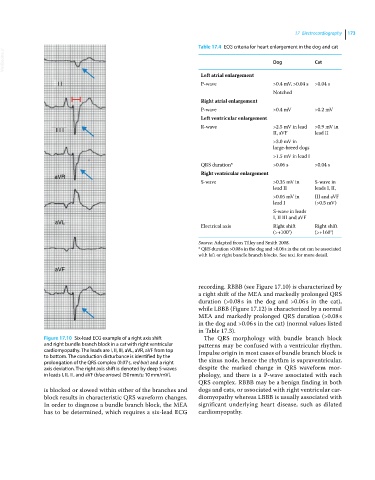
               Figure 17.10  Six‐lead ECG example of a right axis shift   The QRS morphology with bundle branch block
               and right bundle branch block in a cat with right ventricular   patterns may be confused with a ventricular rhythm.
               cardiomyopathy. The leads are I, II, III, aVL, aVR, aVF from top   Impulse origin in most cases of bundle branch block is
               to bottom. The conduction disturbance is identified by the
               prolongation of the QRS complex (0.07 s, red bar) and a right   the sinus node, hence the rhythm is supraventricular,
               axis deviation. The right axis shift is denoted by deep S‐waves   despite the marked change in QRS waveform mor-
               in leads I, II, III, and aVF (blue arrows) (50 mm/s; 10 mm/mV).  phology, and there is a  P‐wave  associated with each
                                                                  QRS complex. RBBB may be a benign finding in both
               is blocked or slowed within either of the branches and   dogs and cats, or associated with right ventricular car-
               block results in characteristic QRS waveform changes.   diomyopathy whereas LBBB is usually associated with
               In order to diagnose a bundle branch block, the MEA   significant underlying heart disease, such as dilated
               has to be determined, which requires a six‐lead ECG   cardiomyopathy.
   200   201   202   203   204   205   206   207   208   209   210