
Page 208 - Clinical Small Animal Internal Medicine
P. 208
176 Section 3 Cardiovascular Disease
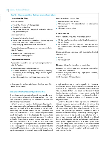
VetBooks.ir Box 18.1 Disease conditions that may produce heart failure Increased resistance to ejection
Impaired cardiac filling
Pericardial disease ● Pulmonic/aortic valve stenosis
Pulmonary/aortic thromboembolism or obstruction
Pericardial effusion with tamponade ●
● (e.g., tumors)
Constrictive pericarditis
● Pulmonary hypertension
Uncommon forms of congenital pericardial disease ●
●
(e.g., pericardial cysts)
Volume overload
Inflow obstructions
Altered blood flow resulting in volume overload
Tricuspid/mitral valve stenosis
●
Uncommon forms of congenital heart disease (e.g., cor ● Valvular insufficiencies (congenital dysplasia, degenera-
●
triatriatum, supravalvular mitral stenosis) tive or infective)
Neoplasia (e.g., obstructive heart base tumors) ● Left to right shunting (e.g., patent ductus arteriosis, ven-
●
tricular septal defect, atrial septal defect, arteriovenous
Myocardial disease that has a primary component of dias- fistulas)
tolic dysfunction
Disease conditions associated with chronically elevated
Hypertrophic cardiomyopathy
● cardiac output
Restrictive cardiomyopathy
●
Anemia
●
Impaired cardiac ejection ● Hyperthryoidism
Myocardial disease that has a primary component of sys-
tolic dysfunction Disorders of impulse formation or conduction
Dilated cardiomyopathy (idiopathic) Sustained tachyarrhythmias (e.g., supraventricular tachy-
●
Ischemic, nutritional (e.g., taurine deficiency), toxic (e.g., cardia, atrial fibrillation)
●
adriamycin), or infectious (e.g., Chagas disease) myocar- Sustained bradyarrhythmias (e.g., high‐grade AV block,
dial disorders sinus bradycardia)
Arrhythmogenic right ventricular cardiomyopathy Wolff–Parkinson–White syndrome
●
actin molecules and myosin heads that is required for systolic performance in cardiac muscle. An alternative
contraction to occur. mechanism, termed length‐dependent activation, seems
to account for augmented ventricular systolic function
with diastolic stretch. The exact mechanisms behind
Determinants of Ventricular Systolic Function
length‐dependent activation remain controversial but
The primary determinants of ventricular systolic func- the basic premise is that up to a maximum length, the
tion are preload, afterload, and contractility. Heart rate myofilaments become sensitized to calcium as sarcomere
and ventricular synchrony are additional factors that length increases.
influence systolic function. The force, tension or stress experienced by the ven-
It has long been recognized that increased cardiac fill- tricular myocytes during contraction is termed the
ing enhances systolic performance. In the normal heart, afterload. Afterload is the degree of interference to ven-
increased preload, which represents the end‐diastolic tricular ejection and is determined primarily by the sys-
distending force of the ventricular wall, produces this temic (or pulmonary) vascular resistance with a
enhanced performance as defined by Starling’s law of the contribution from aortic (or pulmonary arterial) imped-
heart. Surrogates for preload include the end‐diastolic ance. Impedance is dependent on the physical proper-
volume, diameter, or end‐diastolic pressure. Therefore, ties of the vascular wall and the blood (e.g., blood
preload is dependent on venous return, total blood vol- density and viscosity, arterial wall diameter, and viscoe-
ume, and distribution of the blood volume within the lasticity) and represents the ratio of aortic pressure to
vascular system. The recognition that the ascending limb flow. The Anrep effect dictates that an acute increase in
of the Starling curve in skeletal muscle is dictated by the afterload enhances ventricular function via stimulation
extent and “optimization” of actin‐myosin filament over- of sarcolemmal stretch receptors and increased cyto-
lap does not appear to be solely responsible for enhanced solic calcium levels.