
Page 203 - Clinical Small Animal Internal Medicine
P. 203
17 Electrocardiography 171
between 0° and +160°. Calculation of the MEA can be plex is isoelectric (Figure 17.8, step 1), that is, the lead
VetBooks.ir done in several ways. One way is to examine the polar- in which the net polarity of the QRS complex is closest
to zero. (i.e., the positive and negative deflections of the
ity of the QRS complexes from each of the six limb
QRS waves cancel each other out). In some instances,
leads and identify the ECG lead in which the QRS com-
this will be the lead in which the QRS waves are the
smallest and in other instances the lead in which the
aVF – amplitude of the positive R‐wave is the same as the sum
II – –90° III – of the negative deflections (Q‐ and S‐wave, i.e., net
–180° –60° polarity of the QRS).
Then identify the limb lead that is perpendicular to
aVR + aVL + the isoelectric lead on the hexaxial lead system
–150° RA LA –30°
(Figure 17.8, step 2). The MEA will exist in a direction
either towards the positive or negative pole of the per-
pendicular lead, depending on the polarity of the QRS
I– +/–180° 0° I+ complex in that lead (Figure 17.8, step 3). The net polar-
ity of the QRS complex in lead II is positive in the ECG
example in Figure 17.8, so the MEA points to +60°
+150° +30° (Figure 17.8, step 4). This is considered a normal MEA
aVL – RL LL aVR – for a canine.
To obtain a general direction of the MEA, a quick
+120° +60° approach called the “pie method” may be useful
III + +90° II + (Figure 17.9). First, determine the net polarity of the QRS
aVF + complex in lead I and aVF. These are the two leads which
Figure 17.7 The hexaxial or six‐lead ECG system divides the divide the heart into two perpendicular halves and create
frontal plane into 12 segments, similar to the slices of a pie. By four equal pieces (quarters) of a pie.
doing so, the six‐lead ECG examination records the electrical With a normal MEA of depolarization, both lead I and
activity of the heart from six different vantage points along the aVF have a net positive polarity, and the overlapping
frontal plane in 30° increments.
I
aVF –
II – –90° III –
II –180° –60°
3.
aVR + aVL +
RA LA –30°
–150°
III
I– +/–180° 0° I+
aVR
2.
aVL +150° 4. +30°
1. aVL – RL LL aVR –
aVF +120° +60°
III + +90° II +
aVF +
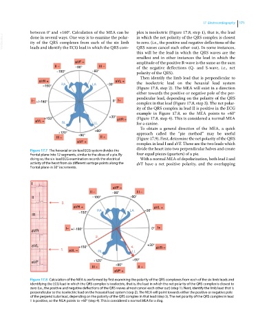
Figure 17.8 Calculation of the MEA is performed by first examining the polarity of the QRS complexes from each of the six limb leads and
identifying the ECG lead in which the QRS complex is isoelectric, that is, the lead in which the net polarity of the QRS complex is closest to
zero (i.e., the positive and negative deflections of the QRS waves almost cancel each other out) (step 1). Next, identify the limb lead that is
perpendicular to the isoelectric lead on the hexaxial lead system (step 2). The MEA will point towards either the positive or negative pole
of the perpendicular lead, depending on the polarity of the QRS complex in that lead (step 3). The net polarity of the QRS complex in lead
II is positive, so the MEA points to +60° (step 4). This is considered a normal MEA for a dog.