Page 206 - Clinical Small Animal Internal Medicine
P. 206
174  Section 3  Cardiovascular Disease


  VetBooks.ir


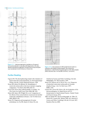
            Figure 17.11  Atrial enlargement is manifest as a P‐wave of
            increased amplitude in a poodle with degenerative AV valve
            disease and atrial enlargement. The blue arrow shows the tall P‐  Figure 17.12  ECG example of a left bundle branch block in a
            wave (amplitude 0.6 mV) (50 mm/s; 10 mm/mV).      rottweiler with dilated cardiomyopathy. The conduction
                                                              disturbance is identified by the prolongation of the QRS complex
                                                              (0.08 s; blue bar) but a normal MEA (50 mm/s; 10 mm/mV).
              Further Reading


            Fogoros RN. The electrophysiology study in the evaluation of   Textbook of Canine and Feline Cardiology, 2nd edn.
              supraventricular tachyarrhythmias. In: Electrophysiologic   Philadelphia, PA: WB Saunders, 1999.
              Testing, 3rd edn. Oxford: Blackwell Science, 1998.  Moise NS, Gilmour RF Jr, Riccio ML, et al. Diagnosis
            Kraus MS, Moise NS, Rishniw M. Morphology of        of inherited ventricular tachycardia in German
              ventricular tachycardia in the boxer and pace mapping   shepherd dogs. J Am Vet Med Assoc 1997;
              comparison. J Vet Intern Med 2002; 16: 153.       210(3): 403.
            Meurs KM. Boxer dog cardiomyopathy: an update. Vet   Oyama MA, Kraus MS, Gelzer AR, eds. Evaluation of the
              Clin North Am Small Anim Pract 2004; 34: 1235.    electrocardiogram. In: Rapid Review of ECG
            Meurs KM, Spier AW, Wright NA, et al. Comparison of   Interpretation in Small Animal Practice. Oxford: Taylor
              the effects of four antiarrhythmic treatments for familial   and Francis Group, 2014.
              ventricular arrhythmias in Boxers. J Am Vet Med Assoc   Tilley LP, Smith WK. Electrocardiography. In: Tilley LP,
              2002; 221(4): 522.                                Smith WK, Oyama MA, Sleeper MM, eds. Manual of
            Moise NS. Diagnosis and management of canine        Canine and Feline Cardiology, 4th edn. St Louis, MO:
              arrhythmias. In: Fox PR, Sisson D, Moise NS, eds.   Saunders Elsevier, 2008.

