
Page 204 - Clinical Small Animal Internal Medicine
P. 204
172 Section 3 Cardiovascular Disease
VetBooks.ir I 1. II – aVF – –60° III –
–90°
–180°
II
4.
aVR + 5. aVL +
III –150° RA LA –30°
aVR I– +/–180° 0° I+
3.
aVL
+30°
+150° aVR –
aVL – RL LL
aVF
2. +60°
+120°
+90°
III + II +
aVF +
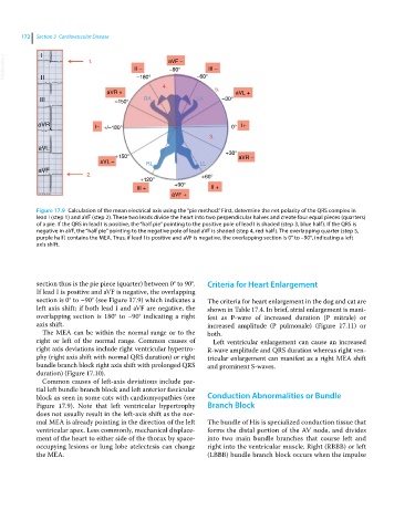
Figure 17.9 Calculation of the mean electrical axis using the “pie method.” First, determine the net polarity of the QRS complex in
lead I (step 1) and aVF (step 2). These two leads divide the heart into two perpendicular halves and create four equal pieces (quarters)
of a pie. If the QRS in lead I is positive, the “half pie” pointing to the positive pole of lead I is shaded (step 3, blue half). If the QRS is
negative in aVF, the “half pie” pointing to the negative pole of lead aVF is shaded (step 4, red half). The overlapping quarter (step 5,
purple half) contains the MEA. Thus, if lead I is positive and aVF is negative, the overlapping section is 0° to –90°, indicating a left
axis shift.
section thus is the pie piece (quarter) between 0° to 90°. Criteria for Heart Enlargement
If lead I is positive and aVF is negative, the overlapping
section is 0° to –90° (see Figure 17.9) which indicates a The criteria for heart enlargement in the dog and cat are
left axis shift; if both lead I and aVF are negative, the shown in Table 17.4. In brief, atrial enlargement is mani-
overlapping section is 180° to –90° indicating a right fest as P‐wave of increased duration (P mitrale) or
axis shift. increased amplitude (P pulmonale) (Figure 17.11) or
The MEA can be within the normal range or to the both.
right or left of the normal range. Common causes of Left ventricular enlargement can cause an increased
right axis deviations include right ventricular hypertro- R‐wave amplitude and QRS duration whereas right ven-
phy (right axis shift with normal QRS duration) or right tricular enlargement can manifest as a right MEA shift
bundle branch block right axis shift with prolonged QRS and prominent S‐waves.
duration) (Figure 17.10).
Common causes of left‐axis deviations include par-
tial left bundle branch block and left anterior fascicular
block as seen in some cats with cardiomyopathies (see Conduction Abnormalities or Bundle
Figure 17.9). Note that left ventricular hypertrophy Branch Block
does not usually result in the left‐axis shift as the nor-
mal MEA is already pointing in the direction of the left The bundle of His is specialized conduction tissue that
ventricular apex. Less commonly, mechanical displace- forms the distal portion of the AV node, and divides
ment of the heart to either side of the thorax by space‐ into two main bundle branches that course left and
occupying lesions or lung lobe atelectesis can change right into the ventricular muscle. Right (RBBB) or left
the MEA. (LBBB) bundle branch block occurs when the impulse