
Page 201 - Clinical Small Animal Internal Medicine
P. 201
17 Electrocardiography 169
ECG sensitivity refers to the amplitude of the wave-
Box 17.1 Summary of ECG waveforms
VetBooks.ir ● The P‐wave represents atrial depolarization forms based on electrical voltage. At standard sensitivity,
an impulse of 1 millivolt (mV) will inscribe a waveform
The Q‐wave is the first negative deflection represent-
●
voltages are normally low (i.e., in cats), sensitivity can be
ing ventricular septal depolarization amplitude that is 10 mm tall. In cases where the ECG
The R‐wave is the first positive deflection representing increased (doubled) so that an impulse of 0.5 mV will
●
early ventricular depolarization create a waveform of 10 mm amplitude, thereby increas-
The S‐wave is the next negative deflection represent- ing the ability to visually inspect the individual ECG
●
ing late ventricular depolarization waveforms. Conversely, in animals with severe heart
The ST segment/T‐wave represents ventricular enlargement, sensitivity can be decreased (halved) so
●
repolarization that waveforms do not extend past the borders of the
recording paper.
ment. In order to assess these characteristics, one must
understand how paper speed and sensitivity settings Heart Rate
affect ECG interpretation. If the heart rhythm is regular, an “instantaneous” heart
rate is easy to calculate: count the number of 1 mm
squares (little boxes) between two heart beats (RR inter-
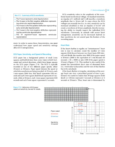
ECG Paper, Sensitivity, and Speed of Recording
val) and divide this number into 3000 if the paper speed
ECG paper has a background pattern of small 1 mm is 50 mm/s (3000 is the equivalent of 1 min because 60
squares and bold division lines every 5 mm in both hori- seconds × 50 = 3000) or into 1500 if the paper speed is
zontal and vertical directions, which form larger squares 25 mm/s (Figure 17.5). This method is also useful if the
on the ECG paper (Figure 17.4). The ECG is typically animal has intermittent short runs of abnormal beats
recorded at one of two different paper speeds, either (i.e., ventricular tachycardia) and the rate of that rhythm
25 mm/s or 50 mm/s. Paper speed denotes the speed at has to be calculated.
which the waveforms are being recorded. At 25 mm/s, each If the heart rhythm is irregular, calculation of the aver-
1 mm square (little box, thin lined) represents 0.04 sec- age heart rate over a prescribed period of time is per-
onds and each 5 mm square (bold lined) represents 0.2 sec- formed. It is useful to realize that 30 large squares (bold
onds, while at 50 mm/s, each 1 mm square represents 0.02 lined) or 150 mm represent 3 seconds at 50 mm/s or 6
seconds and each 5 mm square represents 0.1 seconds. seconds at 25 mm/s. Thus, heart rate is determined by
Figure 17.4 Calibration of ECG paper
speed and sensitivity. See text for details.
0.2sec
0.1sec
0.04sec
Paper speed: 0.02sec
25mm/sec
1mV
50mm/sec
0.1mV
Sensitivity:
Amplitude
10mm/mV
Time