
Page 193 - Clinical Small Animal Internal Medicine
P. 193
16 Imaging in Cardiovascular Disease 161
VetBooks.ir (a)
Width = 15 mm
Length = 35 mm
(b)
(c) (d)
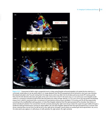
Figure 16.31 Assessment of left‐to‐right congenital shunts in dogs using Doppler echocardiography (a,b: patent ductus arteriosus; c:
ventricular septal defect; d: atrial septal defect). (a) Image obtained from the left parasternal cranial transaortic short‐axis view showing
the aliased turbulent color flow jet coming from the large ductus arteriosus (DA) into the dilated pulmonary trunk (PT) during systole.
Note that the DA diameter decreases abruptly at the point of insertion in the PT. (b) (same dog as in a) Continuous‐wave Doppler mode
confirms a continuous turbulent positive flow into the pulmonary artery of high velocity ( 5.3 m/s and 3.8 m/s during systole and diastole,
respectively), which is consistent with a normal aortic‐to‐pulmonary artery pressure gradient ( 112 mmHg and 58 mmHg, respectively,
according to the modified Bernoulli equation). (c) Color flow Doppler obtained from the right parasternal five‐chamber view shows an
abnormal aliased turbulent flow (arrow) from the left ventricle entering the right ventricle just below the septal tricuspid leaflet, through a
moderate sized perimembranous ventricular septal defect. (d) Color flow Doppler obtained from the right parasternal four‐chamber view
shows a laminar flow (arrow) from the left atrium to the right atrium through a small ostium secundumtype atrial septal defect. Ao, aorta;
IVS, interventricular septum; LA, left atrium; LV, left ventricle; RA, right atrium. RV, right ventricle.