
Page 194 - Clinical Small Animal Internal Medicine
P. 194
162 Section 3 Cardiovascular Disease
VetBooks.ir Radial motion Tissue Doppler Imaging Longitudinal motion
(myocardial velocity, m/s)
Strain rate Imaging
–1
(velocity of myocardial deformation, s )
Strain Imaging
(myocardial deformation, %)
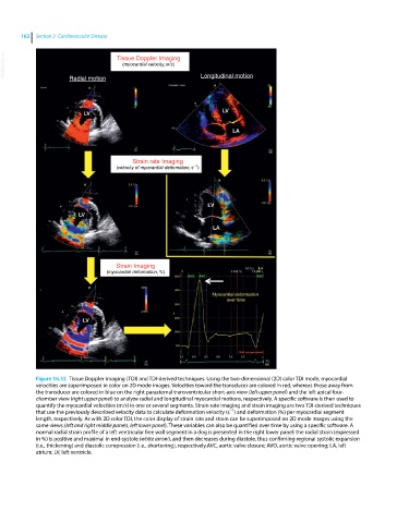
Figure 16.32 Tissue Doppler imaging (TDI) and TDI‐derived techniques. Using the two‐dimensional (2D) color TDI mode, myocardial
velocities are superimposed in color on 2D mode images. Velocities toward the transducer are colored in red, whereas those away from
the transducer are colored in blue on the right parasternal transventricular short‐axis view (left upper panel) and the left apical four‐
chamber view (right upper panel) to analyze radial and longitudinal myocardial motions, respectively. A specific software is then used to
quantify the myocardial velocities (m/s) in one or several segments. Strain rate imaging and strain imaging are two TDI‐derived techniques
−1
that use the previously described velocity data to calculate deformation velocity (s ) and deformation (%) per myocardial segment
length, respectively. As with 2D color TDI, the color display of strain rate and strain can be superimposed on 2D mode images using the
same views (left and right middle panels, left lower panel). These variables can also be quantified over time by using a specific software. A
normal radial strain profile of a left ventricular free wall segment in a dog is presented in the right lower panel: the radial strain (expressed
in %) is positive and maximal in end‐systole (white arrow), and then decreases during diastole, thus confirming regional systolic expansion
(i.e., thickening) and diastolic compression (i.e., shortening), respectively.AVC, aortic valve closure; AVO, aortic valve opening; LA, left
atrium; LV, left ventricle.