
Page 287 - Clinical Small Animal Internal Medicine
P. 287
26 Canine Myocardial Disease 255
of pink‐tinged fluid may be noted. Pleural effusion from recording (24‐hour ambulatory ECG) is the most appro-
VetBooks.ir right‐sided CHF can also produce increased respiratory priate form of ECG in breeds where ventricular arrhyth-
mias are common or an early finding such as Dobermans,
rate and effort. Other signs include exercise intolerance,
weakness, syncope, abdominal distension due to ascites,
fibrillation, or if a cause for syncope is sought. In cases
weight loss, and reduced appetite or anorexia (see for longer term heart rate (HR) assessment in atrial
Chapter 15). Sometimes SD is the first clinical sign noted. where echocardiography is not immediately available,
The typical clinical signs exhibited may vary by breed. thoracic radiographs may be useful for detecting cardio-
For example, Dobermans tend to have a higher incidence megaly, and plasma N‐terminal pro‐B‐type natriuretic
of syncope and SD and their pulmonary edema and peptide (NT‐proBNP) level can be useful in Dobermans
resultant respiratory signs are often quite severe, whereas for assessing likelihood of preclinical disease. For dogs
IWs and Newfoundlands frequently present with evi- with suspected CHF, thoracic radiographs and serum
dence of right‐sided CHF (ascites and pleural effusion). biochemistry should be performed. The role for blood‐
The most common abnormalities detected on physical based tests including biomarkers, taurine levels, and
examination are a soft systolic murmur over the mitral or genetic tests is also discussed below. Screening should be
tricuspid area (often grade 1–3/6) and weak peripheral performed yearly in high‐risk breeds.
pulses. However, various studies have reported murmurs
in as few as 33% and as many as 76%, so the absence of a Electrocardiography
murmur does not rule DCM out, and physical examina- While arrhythmias are frequently reported on ECG
tion may be normal in the early preclinical phase. (89% of cases in one large retrospective study), they can
Additional findings may include a diastolic gallop (low‐ be very intermittent. Ventricular arrhythmias (VA)
frequency third heart sound), an arrhythmia (ventricular (ventricular premature contractions [VPCs] or ventricu-
premature contractions and atrial fibrillation most lar tachycardia [VT]) and atrial fibrillation (AF) are most
common), pulse deficits (if an arrhythmia is present), common, but are not specific for DCM on their own
and jugular venous distension or pulsation. For dogs in (Figures 26.1 and 26.2).
CHF, additional findings may include pale or cyanotic There are differences between breeds in arrhythmia
mucous membranes, increased lung sounds or pulmo- manifestation. VA are very common in Dobermans, box-
nary crackles, tachycardia, tachypnea, dyspnea, hypo- ers, and Great Danes, so their presence should always
thermia, and hepatomegaly (hepatic congestion) or a raise suspicion of DCM in these breeds. In one study in
fluid wave (ascites) on abdominal palpation. Dobermans, one or more VPCs on a 5‐min ECG was
almost 97% specific for detecting a level of VA on Holter
recording that would be suggestive of DCM. However,
Diagnosis
the presence of VPCs on a 5‐min ECG alone is fairly
The diagnosis of DCM involves a minimum of an insensitive for detecting DCM. Studies report 44–64% of
echocardiogram and electrocardiogram (ECG). Holter affected Dobermans and 54% of affected Great Danes
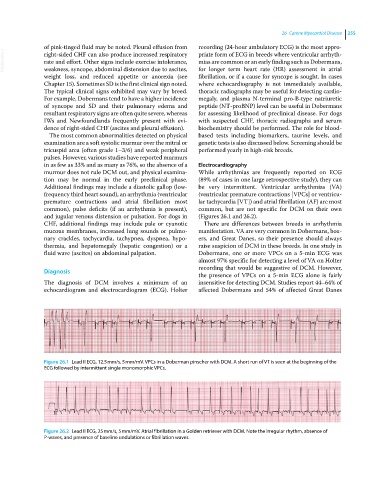
Figure 26.1 Lead II ECG, 12.5 mm/s, 5 mm/mV. VPCs in a Doberman pinscher with DCM. A short run of VT is seen at the beginning of the
ECG followed by intermittent single monomorphic VPCs.
Figure 26.2 Lead II ECG, 25 mm/s, 5 mm/mV. Atrial fibrillation in a Golden retriever with DCM. Note the irregular rhythm, absence of
P‐waves, and presence of baseline undulations or fibrillation waves.