
Page 289 - Clinical Small Animal Internal Medicine
P. 289
26 Canine Myocardial Disease 257
face of severe dilation on echocardiography. This Linear dimensions (diameters) are generated from an M‐
VetBooks.ir phenomenon might be due to the Doberman’s relatively mode of the LV in a short‐ or long‐axis view immediately
below the mitral valve, and volumes are preferentially
deep chest conformation and the relative lack of right
heart involvement compared to other breeds. Breeds
asternal long‐axis or left apical four‐chamber view and
with frequent right‐sided involvement such as IWs and generated by tracing the LV endocardium in a right par-
Newfoundlands often also have pleural effusion, as do using automated software for Simpson’s method of discs.
dogs that develop AF. These parameters are often indexed to body weight (BW)
or body surface area (BSA) to normalize for differences in
Echocardiography body size and conformation. In Dobermans, volume
Echocardiography is necessary for identification and indices have been shown to be more sensitive in detec-
quantification of the characteristic structural abnormali- tion of LV dilation than diameters. Normal parameters
ties, including LV chamber dilation and systolic dysfunc- and breed‐specific criteria suggesting the diagnosis of
tion, and equally important for ruling out other cardiac DCM from various studies are presented in Table 26.1.
causes apart from DCM for these findings (e.g., valvular The reader is referred to the Further Reading list for more
disease, congenital shunt) (Figure 26.4). detailed information on the methods and views used.
Increased LV end‐systolic dimension (LVESD) and/or Left ventricle wall motion is globally and symmetri-
volume (LVESV) is noted, along with increased LV end‐ cally reduced in most cases, but some dogs may have
diastolic dimension (LVEDD) and/or volume (LVEDV). increased septal motion compared to the free wall owing
(a) (b)
(c) (d)
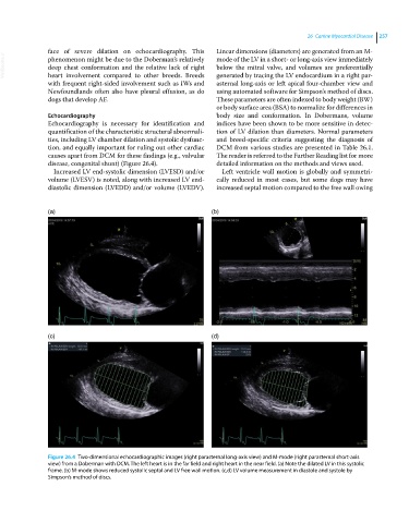
Figure 26.4 Two‐dimensional echocardiographic images (right parasternal long‐axis view) and M‐mode (right parasternal short‐axis
view) from a Doberman with DCM. The left heart is in the far field and right heart in the near field. (a) Note the dilated LV in this systolic
frame. (b) M‐mode shows reduced systolic septal and LV free wall motion. (c,d) LV volume measurement in diastole and systole by
Simpson’s method of discs.