
Page 290 - Clinical Small Animal Internal Medicine
P. 290
258 Section 3 Cardiovascular Disease
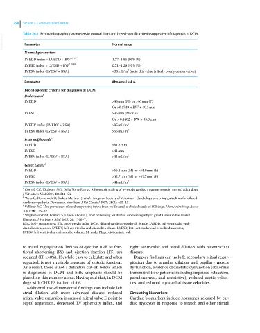
VetBooks.ir Table 26.1 Echocardiographic parameters in normal dogs and breed‐specific criteria suggestive of diagnosis of DCM
Parameter
Normal value
Normal parameters
LVEDD index = LVEDD ÷ BW 0.294 a 1.27–1.85 (95% PI)
LVESD index = LVESD ÷ BW 0.315 a 0.71–1.26 (95% PI)
2
LVESV index (LVESV ÷ BSA) <30 mL/m (note this value is likely overly conservative)
Parameter Abnormal value
Breed‐specific criteria for diagnosis of DCM
Dobermans b
LVEDD >48 mm (M) or >46 mm (F)
Or >0.1749 × BW + 40.3 mm
LVESD >36 mm (M or F)
Or > 0.1402 × BW + 35.0 mm
LVEDV index (LVEDV ÷ BSA) >95 mL/m 2
LVESV index (LVESV ÷ BSA) >55 mL/m 2
c
Irish wolfhounds
LVEDD >61.2 mm
LVESD >41 mm
LVESV index (LVESV ÷ BSA) >41 mL/m 2
d
Great Danes
LVEDD >56.1 mm (M) or >54.0 mm (F)
LVESD >42.7 mm (M) or >41.7 mm (F)
LVESV index (LVESV ÷ BSA) >44 mL/m 2
a Cornell CC, Kittleson MD, Della Torre P, et al. Allomettric scaling of M‐mode cardiac measurements in normal adult dogs.
J Vet Intern Med 2004; 18: 311–21.
b Wess G, Domenech O, Dukes‐McEwan J, et al. European Society of Veterinary Cardiology screening guidelines for dilated
cardiomyopathy in Doberman pinschers. J Vet Cardiol 2017; 19(5): 405–15.
c Vollmar AC. The prevalence of cardiomyopathy in the Irish wolfhound: a clinical study of 500 dogs. J Am Anim Hosp Assoc
2000; 36: 125–32.
d Stephenson HM, Fonfara S, López‐Alvarez J, et al. Screening for dilated cardiomyopathy in great Danes in the United
Kingdom. J Vet Intern Med 2012; 26: 1140–7.
BSA, body surface area; BW, body weight in kg; DCM, dilated cardiomyopathy; F, female; LVEDD, left ventricular end‐
diastolic dimension; LVEDV, left ventricular end‐diastolic volume; LVESD, left ventricular end‐systolic dimension;
LVESV, left ventricular end‐systolic volume; M, male; PI, prediction interval.
to mitral regurgitation. Indices of ejection such as frac- right ventricular and atrial dilation with biventricular
tional shortening (FS) and ejection fraction (EF) are disease.
reduced (EF <40%). FS, while easy to calculate and often Doppler findings can include secondary mitral regur-
reported, is not a reliable measure of systolic function. gitation due to annulus dilation and papillary muscle
As a result, there is not a definitive cut‐off below which dysfunction, evidence of diastolic dysfunction (abnormal
is diagnostic of DCM and little emphasis should be transmitral flow patterns including impaired relaxation,
placed on this number alone. Having said that, in DCM pseudonormal, and restrictive), reduced aortic veloci-
dogs with CHF, FS is often <15%. ties, and reduced myocardial tissue velocities.
Additional two‐dimensional findings can include left
atrial dilation with more advanced disease, reduced Circulating Biomarkers
mitral valve excursion, increased mitral valve E‐point to Cardiac biomarkers include hormones released by car-
septal separation, decreased LV sphericity index, and diac myocytes in response to stretch and other stimuli