
Page 460 - Feline diagnostic imaging
P. 460
472 27 Urinary Disease
(a) (b)
(d)
(c)
(e)
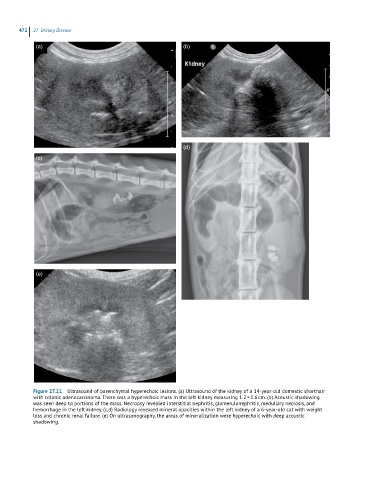
Figure 27.21 Ultrasound of parenchymal hyperechoic lesions. (a) Ultrasound of the kidney of a 14-year-old domestic shorthair
with colonic adenocarcinoma. There was a hyperechoic mass in the left kidney measuring 1.2 × 1.6 cm. (b) Acoustic shadowing
was seen deep to portions of the mass. Necropsy revealed interstitial nephritis, glomerulonephritis, medullary necrosis, and
hemorrhage in the left kidney. (c,d) Radiology revealed mineral opacities within the left kidney of a 6-year-old cat with weight
loss and chronic renal failure. (e) On ultrasonography, the areas of mineralization were hyperechoic with deep acoustic
shadowing.