
Page 462 - Feline diagnostic imaging
P. 462
(a) (b)
(c)
(d)
(e)
(f)
(g)
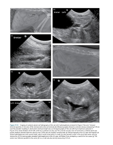
Figure 27.23 Imaging of ureteral calculi. (a) Radiography of the cat with hydronephrosis pictured in Figure 27.6e and f showed
mineral opacities in the area of the kidneys and ureter (arrowhead). (b) Ultrasonography showed a ureteral calculus measuring 0.39 cm
near the bladder resulting in obstruction and hydronephrosis. (c) Ultrasonography of the cat with hydronephrosis pictured in
Figure 27.6c shows dilation of the left ureter by a ureteral calculus. (d) The ureteral calculus was removed and a ureteral stent was
placed. Analysis showed that the calculus was 100% calcium oxalate monohydrate. (e) Ultrasonography of an 11-year-old Ragdoll cat
shows hydronephrosis with several small calculi in the renal pelvis. A small calculus (arrow) is seen in the proximal ureter causing
obstruction. (f) Ultrasonography revealed hydronephrosis in this 16-year-old Maine Coon, prompting a search for the cause. (g) The
ureter was followed parallel to the aorta (A) to a small calculus (arrow) measuring 0.23 cm.