
Page 466 - Feline diagnostic imaging
P. 466
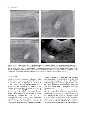
478  27  Urinary Disease

                (a)                                       (b)


                (c)                                       (d)


            Figure 27.26  (a) Lateral projection of the caudal abdomen of a 7-year-old Ragdoll with chronic cystitis of more than eight months
            duration. The cranial aspect of the urinary bladder has a pointed appearance consistent with a urachal diverticulum. (b) An excretory
            urogram was performed. During the cystogram phase of the excretory urogram, calculi (small arrows) are seen as filling defects in the
            contrast within the bladder. Notice that the bladder wall is thickened and the urachal diverticulum is visible. (c) At 45 minutes, margins
            of the contrast in the urinary bladder are irregular. (d) Ultrasound image of one of the calculi measured at 0.93 cm.


            27.4.3  Cystitis
                                                              resulting  from  trauma  five  years  earlier  and   prolonged
            Cystitis  is  not  apparent  on  survey  radiography  unless   antibiotic  therapy  were  considered  to  be  predisposing
            there is increased or decreased opacity in the area of the   causes. On ultrasonography, the bladder wall was thick-
            bladder.  Gas  in  the  wall,  ligaments,  or  lumen  of  the   ened and irregular. It was mainly hypoechoic but there
              urinary  bladder  indicates  emphysematous  cystitis   was  a  thick  hyperechoic  mucosal  surface.  Postmortem
            (Figure 27.30), which is most likely to be associated with   examination revealed a thick granular crust adhering to
            diabetes mellitus although it has been reported in a cat   the bladder wall.
            caused by Escherichia coli [38]. The presence of gas could   Double contrast cystography can help detect cystitis
            also be iatrogenic due to prior catheterization or cysto-  when  the  bladder  wall  is  thickened  (Figure  27.31).
            centesis.  Mineralization  in  the  bladder  is  usually   Positive contrast coats the mucosal border of the blad-
              associated  with  neoplasia  but  in  rare  cases  can  be   der, allowing detection of focal or uniform thickening
              associated  with  cystitis.  Increased  opacity  attributed  to   of the wall adjacent to the air‐filled lumen. The bladder
              mineralization was seen in the urinary bladder of a cat   wall in patients with chronic cystitis is not as distensi-
            with an unusual case of encrusting cystitis  secondary to   ble as a normal bladder. It is better to do multiple test
            Corynebacterium urealyticum [39]. Neurologic problems   injections than to rupture the bladder. The mucosa may