
Page 461 - Feline diagnostic imaging
P. 461
27.3 Ureteral Strictures 473
proportions have changed, possibly due to dietary
changes that acidified the urine but that had the side
effect of releasing osseous calcium carbonate, thereby
promoting hypercalciuria [29]. In one urinary stone labo-
ratory, 10% of calculi submitted consisted of urate or
urate mixed with struvite or calcium oxalate [30]. Only a
small percentage of these cats, usually younger cats,
were confirmed to have a portosystemic shunt. The
majority of the other cats did not have clinical signs of
liver disease and consequently, the possibility of porto-
systemic shunt was not investigated. Xanthine calculi are
relatively rare and may be associated with a congenital
defect in xanthine dehydrogenase [31].
There is an impression that ureterolithiasis is becoming
more common although it is difficult to determine whether
this is an actual increase, there is an increased awareness
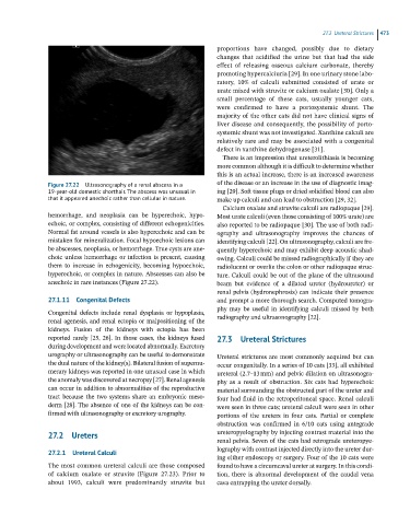
Figure 27.22 Ultrasonography of a renal abscess in a of the disease or an increase in the use of diagnostic imag-
19-year-old domestic shorthair. The abscess was unusual in ing [29]. Soft tissue plugs or dried solidified blood can also
that it appeared anechoic rather than cellular in nature. make up calculi and can lead to obstruction [29, 32].
Calcium oxalate and struvite calculi are radiopaque [29].
hemorrhage, and neoplasia can be hyperechoic, hypo- Most urate calculi (even those consisting of 100% urate) are
echoic, or complex, consisting of different echogenicities. also reported to be radiopaque [30]. The use of both radi-
Normal fat around vessels is also hyperechoic and can be ography and ultrasonography improves the chances of
mistaken for mineralization. Focal hypoechoic lesions can identifying calculi [22]. On ultrasonography, calculi are fre-
be abscesses, neoplasia, or hemorrhage. True cysts are ane- quently hyperechoic and may exhibit deep acoustic shad-
choic unless hemorrhage or infection is present, causing owing. Calculi could be missed radiographically if they are
them to increase in echogenicity, becoming hypoechoic, radiolucent or overlie the colon or other radiopaque struc-
hyperechoic, or complex in nature. Abscesses can also be ture. Calculi could be out of the plane of the ultrasound
anechoic in rare instances (Figure 27.22). beam but evidence of a dilated ureter (hydroureter) or
renal pelvis (hydronephrosis) can indicate their presence
27.1.11 Congenital Defects and prompt a more thorough search. Computed tomogra-
phy may be useful in identifying calculi missed by both
Congenital defects include renal dysplasia or hypoplasia, radiography and ultrasonography [22].
renal agenesis, and renal ectopia or malpositioning of the
kidneys. Fusion of the kidneys with ectopia has been
reported rarely [25, 26]. In those cases, the kidneys fused 27.3 Ureteral Strictures
during development and were located abnormally. Excretory
urography or ultrasonography can be useful to demonstrate Ureteral strictures are most commonly acquired but can
the dual nature of the kidney(s). Bilateral fusion of supernu- occur congenitally. In a series of 10 cats [33], all exhibited
merary kidneys was reported in one unusual case in which ureteral (2.7–13 mm) and pelvic dilation on ultrasonogra-
the anomaly was discovered at necropsy [27]. Renal agenesis phy as a result of obstruction. Six cats had hyperechoic
can occur in addition to abnormalities of the reproductive material surrounding the obstructed part of the ureter and
tract because the two systems share an embryonic meso- four had fluid in the retroperitoneal space. Renal calculi
derm [28]. The absence of one of the kidneys can be con- were seen in three cats; ureteral calculi were seen in other
firmed with ultrasonography or excretory urography. portions of the ureters in four cats. Partial or complete
obstruction was confirmed in 6/10 cats using antegrade
27.2 Ureters ureteropyelography by injecting contrast material into the
renal pelvis. Seven of the cats had retrograde ureteropye-
lography with contrast injected directly into the ureter dur-
27.2.1 Ureteral Calculi
ing either endoscopy or surgery. Four of the 10 cats were
The most common ureteral calculi are those composed found to have a circumcaval ureter at surgery. In this condi-
of calcium oxalate or struvite (Figure 27.23). Prior to tion, there is abnormal development of the caudal vena
about 1993, calculi were predominantly struvite but cava entrapping the ureter dorsally.