
Page 465 - Feline diagnostic imaging
P. 465
27.4 ladder 477
(a) (b) (c)
(d)
(e) (f)
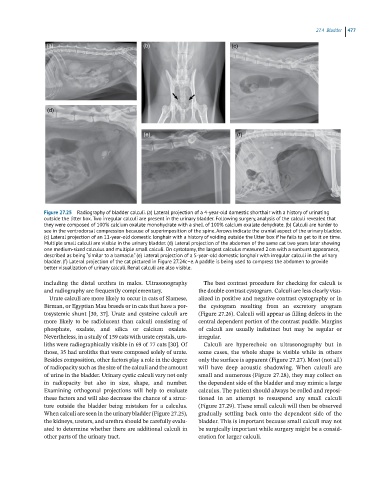
Figure 27.25 Radiography of bladder calculi. (a) Lateral projection of a 4-year-old domestic shorthair with a history of urinating
outside the litter box. Two irregular calculi are present in the urinary bladder. Following surgery, analysis of the calculi revealed that
they were composed of 100% calcium oxalate monohydrate with a shell of 100% calcium oxalate dehydrate. (b) Calculi are harder to
see in the ventrodorsal compression because of superimposition of the spine. Arrows indicate the cranial aspect of the urinary bladder.
(c) Lateral projection of an 11-year-old domestic longhair with a history of voiding outside the litter box if he fails to get to it on time.
Multiple small calculi are visible in the urinary bladder. (d) Lateral projection of the abdomen of the same cat two years later showing
one medium-sized calculus and multiple small calculi. On cystotomy, the largest calculus measured 2 cm with a sunburst appearance,
described as being “similar to a barnacle.” (e) Lateral projection of a 5-year-old domestic longhair with irregular calculi in the urinary
bladder. (f) Lateral projection of the cat pictured in Figure 27.24c–e. A paddle is being used to compress the abdomen to provide
better visualization of urinary calculi. Renal calculi are also visible.
including the distal urethra in males. Ultrasonography The best contrast procedure for checking for calculi is
and radiography are frequently complementary. the double contrast cystogram. Calculi are less clearly visu-
Urate calculi are more likely to occur in cats of Siamese, alized in positive and negative contrast cystography or in
Birman, or Egyptian Mau breeds or in cats that have a por- the cystogram resulting from an excretory urogram
tosystemic shunt [30, 37]. Urate and cysteine calculi are (Figure 27.26). Calculi will appear as filling defects in the
more likely to be radiolucent than calculi consisting of central dependent portion of the contrast puddle. Margins
phosphate, oxalate, and silica or calcium oxalate. of calculi are usually indistinct but may be regular or
Nevertheless, in a study of 159 cats with urate crystals, uro- irregular.
liths were radiographically visible in 69 of 77 cats [30]. Of Calculi are hyperechoic on ultrasonography but in
those, 35 had uroliths that were composed solely of urate. some cases, the whole shape is visible while in others
Besides composition, other factors play a role in the degree only the surface is apparent (Figure 27.27). Most (not all)
of radiopacity such as the size of the calculi and the amount will have deep acoustic shadowing. When calculi are
of urine in the bladder. Urinary cystic calculi vary not only small and numerous (Figure 27.28), they may collect on
in radiopacity but also in size, shape, and number. the dependent side of the bladder and may mimic a large
Examining orthogonal projections will help to evaluate calculus. The patient should always be rolled and reposi-
these factors and will also decrease the chance of a struc- tioned in an attempt to resuspend any small calculi
ture outside the bladder being mistaken for a calculus. (Figure 27.29). These small calculi will then be observed
When calculi are seen in the urinary bladder (Figure 27.25), gradually settling back onto the dependent side of the
the kidneys, ureters, and urethra should be carefully evalu- bladder. This is important because small calculi may not
ated to determine whether there are additional calculi in be surgically important while surgery might be a consid-
other parts of the urinary tract. eration for larger calculi.